 Movie
Movie Controller

[English] 日本語
 Yorodumi
- PDB-2fus: MUTATIONS OF FUMARASE THAT DISTINGUISH BETWEEN THE ACTIVE SITE AN... -
+ Open data

- Basic information
| Entry | Database: PDB / ID: 2fus | ||||||
|---|---|---|---|---|---|---|---|
| Title | MUTATIONS OF FUMARASE THAT DISTINGUISH BETWEEN THE ACTIVE SITE AND A NEARBY DICARBOXYLIC ACID BINDING SITE | ||||||
 Components Components | FUMARASE C | ||||||
 Keywords Keywords | HYDROLYASE / CARBON OXYGEN LYASE / KREB'S CYCLE ENZYME / FUMARATE HYDRATASE | ||||||
| Function / homology |  Function and homology information Function and homology informationfumarate hydratase activity / fumarate hydratase / fumarate metabolic process / malate metabolic process / tricarboxylic acid cycle / response to oxidative stress / identical protein binding / cytoplasm Similarity search - Function | ||||||
| Biological species |  | ||||||
| Method |  X-RAY DIFFRACTION / MOLECULAR REPLACEMENT / Resolution: 2.2 Å X-RAY DIFFRACTION / MOLECULAR REPLACEMENT / Resolution: 2.2 Å | ||||||
 Authors Authors | Weaver, T.M. / Lees, M. / Banaszak, L.J. | ||||||
 Citation Citation | Journal: Protein Sci. / Year: 1997 Title: Mutations of fumarase that distinguish between the active site and a nearby dicarboxylic acid binding site. Authors: Weaver, T. / Lees, M. / Banaszak, L. | ||||||
| History |
|
- Structure visualization
| Structure viewer | Molecule: MolmilJmol/JSmol |
|---|
- Downloads & links
-Download
| PDBx/mmCIF format | 2fus.cif.gz | 233.9 KB | Display | PDBx/mmCIF format |
|---|---|---|---|---|
| PDB format | pdb2fus.ent.gz | 190.6 KB | Display | PDB format |
| PDBx/mmJSON format | 2fus.json.gz | Tree view | PDBx/mmJSON format | |
| Others |  Other downloads Other downloads |
-Validation report
| Arichive directory | https://data.pdbj.org/pub/pdb/validation_reports/fu/2fusftp://data.pdbj.org/pub/pdb/validation_reports/fu/2fus | HTTPS FTP |
|---|
-Related structure data
| Related structure data |  1furC  1fuoS S: Starting model for refinement C: citing same article ( |
|---|---|
| Similar structure data |


-Links
 PDBj
PDBj

- Assembly
| Deposited unit | 
| ||||||||
|---|---|---|---|---|---|---|---|---|---|
| 1 | 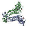
| ||||||||
| Unit cell |
| ||||||||
| Noncrystallographic symmetry (NCS) | NCS oper: (Code: given Matrix: (0.4695, -0.0031, 0.8829), Vector: |
-Components
| #1: Protein | Mass: 50524.730 Da / Num. of mol.: 2 / Mutation: H129N Source method: isolated from a genetically manipulated source Source: (gene. exp.) #2: Chemical |   Mass: 192.124 Da / Num. of mol.: 2 / Source method: obtained synthetically / Formula: C6H8O7 Mass: 192.124 Da / Num. of mol.: 2 / Source method: obtained synthetically / Formula: C6H8O7#3: Water | ChemComp-HOH / |  Mass: 18.015 Da / Num. of mol.: 371 / Source method: isolated from a natural source / Formula: H2O Mass: 18.015 Da / Num. of mol.: 371 / Source method: isolated from a natural source / Formula: H2OCompound details | THE SECOND ANION BINDING SITE LOCATED BETWEEN ARG 126 THROUGH ASN 135 HAS BEEN STERICALLY BLOCKED ...THE SECOND ANION BINDING SITE LOCATED BETWEEN ARG 126 THROUGH ASN 135 HAS BEEN STERICALLY | |
|---|
-Experimental details
-Experiment
| Experiment | Method: X-RAY DIFFRACTION / Number of used crystals: 1 |
|---|
- Sample preparation
| Crystal | Density Matthews: 2.46 Å3/Da / Density % sol: 50 % | |||||||||||||||
|---|---|---|---|---|---|---|---|---|---|---|---|---|---|---|---|---|
| Crystal grow | pH: 5 Details: PROTEIN WAS CRYSTALLIZED FROM 150MM CITRATE PH 5.0 AND 14% PEG 4000 | |||||||||||||||
| Crystal grow | *PLUS pH: 6 / Method: unknown | |||||||||||||||
| Components of the solutions | *PLUS
|
-Data collection
| Diffraction | Mean temperature: 298 K |
|---|---|
| Diffraction source | Source: ROTATING ANODE / Type: RIGAKU RUH2R / Wavelength: 1.5418 |
| Detector | Type: SIEMENS / Detector: AREA DETECTOR / Date: Feb 1, 1996 |
| Radiation | Monochromator: GRAPHITE(002) / Monochromatic (M) / Laue (L): M / Scattering type: x-ray |
| Radiation wavelength | Wavelength: 1.5418 Å / Relative weight: 1 |
| Reflection | Resolution: 1.98→8 Å / Num. obs: 60762 / % possible obs: 88 % / Observed criterion σ(I): 1 / Redundancy: 4.3 % / Rmerge(I) obs: 0.07 / Rsym value: 0.117 / Net I/σ(I): 7.4 |
| Reflection shell | Resolution: 1.98→2.06 Å / Redundancy: 2.8 % / Rmerge(I) obs: 0.61 / Mean I/σ(I) obs: 0.435 / Rsym value: 0.71 / % possible all: 46 |
| Reflection shell | *PLUS % possible obs: 46 % |
- Processing
| Software |
| ||||||||||||||||||||||||||||||||||||||||||||||||||||||||||||
|---|---|---|---|---|---|---|---|---|---|---|---|---|---|---|---|---|---|---|---|---|---|---|---|---|---|---|---|---|---|---|---|---|---|---|---|---|---|---|---|---|---|---|---|---|---|---|---|---|---|---|---|---|---|---|---|---|---|---|---|---|---|
| Refinement | Method to determine structure: MOLECULAR REPLACEMENT Starting model: PDB ENTRY 1FUO Resolution: 2.2→8 Å / Rfactor Rfree error: 0.003 / Data cutoff high absF: 10000000 / Data cutoff low absF: 0.001 / Cross valid method: THROUGHOUT / σ(F): 1
| ||||||||||||||||||||||||||||||||||||||||||||||||||||||||||||
| Refinement step | Cycle: LAST / Resolution: 2.2→8 Å
| ||||||||||||||||||||||||||||||||||||||||||||||||||||||||||||
| Refine LS restraints |
| ||||||||||||||||||||||||||||||||||||||||||||||||||||||||||||
| LS refinement shell | Resolution: 2.2→2.3 Å / Total num. of bins used: 8 / % reflection obs: 85.26 % | ||||||||||||||||||||||||||||||||||||||||||||||||||||||||||||
| Xplor file |
| ||||||||||||||||||||||||||||||||||||||||||||||||||||||||||||
| Software | *PLUS Name: X-PLOR / Version: 3.1 / Classification: refinement | ||||||||||||||||||||||||||||||||||||||||||||||||||||||||||||
| Refinement | *PLUS | ||||||||||||||||||||||||||||||||||||||||||||||||||||||||||||
| Solvent computation | *PLUS | ||||||||||||||||||||||||||||||||||||||||||||||||||||||||||||
| Displacement parameters | *PLUS | ||||||||||||||||||||||||||||||||||||||||||||||||||||||||||||
| Refine LS restraints | *PLUS
|
