 Movie
Movie Controller

[English] 日本語
 Yorodumi
- PDB-1fur: FUMARASE MUTANT H188N WITH BOUND SUBSTRATE L-MALATE AT PUTATIVE A... -
+ Open data

- Basic information
| Entry | Database: PDB / ID: 1fur | ||||||
|---|---|---|---|---|---|---|---|
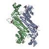
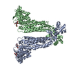
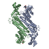
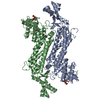
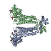
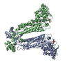
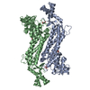
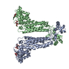
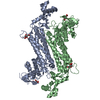
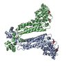
| Title | FUMARASE MUTANT H188N WITH BOUND SUBSTRATE L-MALATE AT PUTATIVE ACTIVATOR SITE | ||||||
 Components Components | FUMARASE C | ||||||
 Keywords Keywords | HYDROLYASE / CARBON OXYGEN LYASE / KREB'S CYCLE ENZYME / FUMARATE HYDRATASE | ||||||
| Function / homology |  Function and homology information Function and homology informationfumarate hydratase activity / fumarate hydratase / fumarate metabolic process / malate metabolic process / tricarboxylic acid cycle / response to oxidative stress / identical protein binding / cytoplasm Similarity search - Function | ||||||
| Biological species |  | ||||||
| Method |  X-RAY DIFFRACTION / MOLECULAR REPLACEMENT / Resolution: 1.95 Å X-RAY DIFFRACTION / MOLECULAR REPLACEMENT / Resolution: 1.95 Å | ||||||
 Authors Authors | Weaver, T.M. / Lees, M. / Banaszak, L.J. | ||||||
 Citation Citation | Journal: Protein Sci. / Year: 1997 Title: Mutations of fumarase that distinguish between the active site and a nearby dicarboxylic acid binding site. Authors: Weaver, T. / Lees, M. / Banaszak, L. | ||||||
| History |
|
- Structure visualization
| Structure viewer | Molecule: MolmilJmol/JSmol |
|---|
- Downloads & links
-Download
| PDBx/mmCIF format | 1fur.cif.gz | 235 KB | Display | PDBx/mmCIF format |
|---|---|---|---|---|
| PDB format | pdb1fur.ent.gz | 191.8 KB | Display | PDB format |
| PDBx/mmJSON format | 1fur.json.gz | Tree view | PDBx/mmJSON format | |
| Others |  Other downloads Other downloads |
-Validation report
| Arichive directory | https://data.pdbj.org/pub/pdb/validation_reports/fu/1furftp://data.pdbj.org/pub/pdb/validation_reports/fu/1fur | HTTPS FTP |
|---|
-Related structure data
| Related structure data |  2fusC  1fuoS S: Starting model for refinement C: citing same article ( |
|---|---|
| Similar structure data |
-Links
 PDBj
PDBj


- Assembly
| Deposited unit | 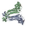
| ||||||||
|---|---|---|---|---|---|---|---|---|---|
| 1 | 
| ||||||||
| Unit cell |
| ||||||||
| Noncrystallographic symmetry (NCS) | NCS oper: (Code: given Matrix: (0.497147, -0.009049, 0.867619), Vector: |
-Components
| #1: Protein | Mass: 50524.727 Da / Num. of mol.: 2 / Mutation: H188N Source method: isolated from a genetically manipulated source Source: (gene. exp.) Description: SITE-DIRECTED MUTANT OF FUMARASE C WAS GENERATED BY PCR. THE NATIVE ENZYME ALSO INCORPORATES A 5 HIS TAG ON THE CARBOXY TERMINUS THAT IS NOT VISIBLE IN THE ELECTRON DENSITY MAPS ...Description: SITE-DIRECTED MUTANT OF FUMARASE C WAS GENERATED BY PCR. THE NATIVE ENZYME ALSO INCORPORATES A 5 HIS TAG ON THE CARBOXY TERMINUS THAT IS NOT VISIBLE IN THE ELECTRON DENSITY MAPS CALCULATED USING PHASES CALCULATED FROM THE FINAL MODEL COORDINATES. Cell line: 293 / Cellular location: CYTOPLASM / Gene: FUMC / Plasmid: PASK40_188EFUMC / Cellular location (production host): CYTOPLASM / Gene (production host): FUMC / Production host: #2: Chemical |   Mass: 134.087 Da / Num. of mol.: 2 / Source method: obtained synthetically / Formula: C4H6O5 Mass: 134.087 Da / Num. of mol.: 2 / Source method: obtained synthetically / Formula: C4H6O5#3: Water | ChemComp-HOH / |  Mass: 18.015 Da / Num. of mol.: 402 / Source method: isolated from a natural source / Formula: H2O Mass: 18.015 Da / Num. of mol.: 402 / Source method: isolated from a natural source / Formula: H2O |
|---|
-Experimental details
-Experiment
| Experiment | Method: X-RAY DIFFRACTION / Number of used crystals: 1 |
|---|
- Sample preparation
| Crystal | Density Matthews: 2.46 Å3/Da / Density % sol: 50 % | |||||||||||||||
|---|---|---|---|---|---|---|---|---|---|---|---|---|---|---|---|---|
| Crystal grow | pH: 5 Details: PROTEIN WAS CRYSTALLIZED FROM 150MM CITRATE PH 5.0 AND 14% PEG 4000 | |||||||||||||||
| Crystal grow | *PLUS pH: 6 / Method: unknown | |||||||||||||||
| Components of the solutions | *PLUS
|
-Data collection
| Diffraction | Mean temperature: 298 K |
|---|---|
| Diffraction source | Source: ROTATING ANODE / Type: RIGAKU RUH2R / Wavelength: 1.5418 |
| Detector | Type: SIEMENS / Detector: AREA DETECTOR / Date: Jul 1, 1996 |
| Radiation | Monochromator: GRAPHITE(002) / Monochromatic (M) / Laue (L): M / Scattering type: x-ray |
| Radiation wavelength | Wavelength: 1.5418 Å / Relative weight: 1 |
| Reflection | Resolution: 1.95→8 Å / Num. obs: 79307 / % possible obs: 88 % / Observed criterion σ(I): 1 / Redundancy: 5.2 % / Rmerge(I) obs: 0.064 / Rsym value: 0.094 / Net I/σ(I): 10.2 |
| Reflection shell | Resolution: 1.81→1.88 Å / Redundancy: 0.75 % / Rmerge(I) obs: 0.36 / Mean I/σ(I) obs: 0.55 / Rsym value: 0.38 / % possible all: 49 |
| Reflection shell | *PLUS % possible obs: 75 % |
- Processing
| Software |
| ||||||||||||||||||||||||||||||||||||||||||||||||||||||||||||
|---|---|---|---|---|---|---|---|---|---|---|---|---|---|---|---|---|---|---|---|---|---|---|---|---|---|---|---|---|---|---|---|---|---|---|---|---|---|---|---|---|---|---|---|---|---|---|---|---|---|---|---|---|---|---|---|---|---|---|---|---|---|
| Refinement | Method to determine structure: MOLECULAR REPLACEMENT Starting model: PDB ENTRY 1FUO Resolution: 1.95→8 Å / Rfactor Rfree error: 0.0025 / Data cutoff high absF: 10000000 / Data cutoff low absF: 0.001 / Cross valid method: THROUGHOUT / σ(F): 1
| ||||||||||||||||||||||||||||||||||||||||||||||||||||||||||||
| Refinement step | Cycle: LAST / Resolution: 1.95→8 Å
| ||||||||||||||||||||||||||||||||||||||||||||||||||||||||||||
| Refine LS restraints |
| ||||||||||||||||||||||||||||||||||||||||||||||||||||||||||||
| Xplor file |
| ||||||||||||||||||||||||||||||||||||||||||||||||||||||||||||
| Software | *PLUS Name: X-PLOR / Version: 3.1 / Classification: refinement | ||||||||||||||||||||||||||||||||||||||||||||||||||||||||||||
| Refinement | *PLUS | ||||||||||||||||||||||||||||||||||||||||||||||||||||||||||||
| Solvent computation | *PLUS | ||||||||||||||||||||||||||||||||||||||||||||||||||||||||||||
| Displacement parameters | *PLUS | ||||||||||||||||||||||||||||||||||||||||||||||||||||||||||||
| Refine LS restraints | *PLUS
|
