 Movie
Movie Controller

[English] 日本語
 Yorodumi
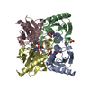
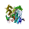
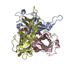
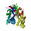
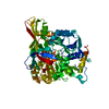
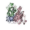
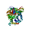
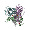
- PDB-2fr6: Crystal Structure of Mouse Cytidine Deaminase Complexed with Cytidine -
+ Open data

- Basic information
| Entry | Database: PDB / ID: 2fr6 | ||||||
|---|---|---|---|---|---|---|---|
| Title | Crystal Structure of Mouse Cytidine Deaminase Complexed with Cytidine | ||||||
 Components Components | Cytidine deaminase | ||||||
 Keywords Keywords | HYDROLASE / CYTIDINE DEAMINASE / ZINC / CYTIDINE / URIDINE / PROTEIN-SUBSTRATE COMPLEX / SUBSTRATE-PRODUCT INTERMEDIATE | ||||||
| Function / homology |  Function and homology information Function and homology informationPyrimidine salvage / CMP catabolic process / dCMP catabolic process / cytidine deaminase / deaminase activity / cellular response to external biotic stimulus / cytidine deaminase activity / UMP salvage / nucleoside binding / Neutrophil degranulation ...Pyrimidine salvage / CMP catabolic process / dCMP catabolic process / cytidine deaminase / deaminase activity / cellular response to external biotic stimulus / cytidine deaminase activity / UMP salvage / nucleoside binding / Neutrophil degranulation / protein homodimerization activity / zinc ion binding / identical protein binding / cytosol Similarity search - Function | ||||||
| Biological species |  | ||||||
| Method |  X-RAY DIFFRACTION / MOLECULAR REPLACEMENT / Resolution: 2.07 Å X-RAY DIFFRACTION / MOLECULAR REPLACEMENT / Resolution: 2.07 Å | ||||||
 Authors Authors | Teh, A.H. | ||||||
 Citation Citation | Journal: Biochemistry / Year: 2006 Title: The 1.48 A Resolution Crystal Structure of the Homotetrameric Cytidine Deaminase from Mouse Authors: Teh, A.H. / Kimura, M. / Yamamoto, M. / Tanaka, N. / Yamaguchi, I. / Kumasaka, T. | ||||||
| History |
|
- Structure visualization
| Structure viewer | Molecule: MolmilJmol/JSmol |
|---|
- Downloads & links
-Download
| PDBx/mmCIF format | 2fr6.cif.gz | 130 KB | Display | PDBx/mmCIF format |
|---|---|---|---|---|
| PDB format | pdb2fr6.ent.gz | 99.9 KB | Display | PDB format |
| PDBx/mmJSON format | 2fr6.json.gz | Tree view | PDBx/mmJSON format | |
| Others |  Other downloads Other downloads |
-Validation report
| Arichive directory | https://data.pdbj.org/pub/pdb/validation_reports/fr/2fr6ftp://data.pdbj.org/pub/pdb/validation_reports/fr/2fr6 | HTTPS FTP |
|---|
-Related structure data
| Related structure data | 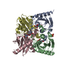 1zabC  2fr5SC S: Starting model for refinement C: citing same article ( |
|---|---|
| Similar structure data |


-Links
 PDBj
PDBj


- Assembly
| Deposited unit | 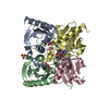
| |||||||||
|---|---|---|---|---|---|---|---|---|---|---|
| 1 |
| |||||||||
| 2 | 
| |||||||||
| 3 | 
| |||||||||
| Unit cell |
| |||||||||
| Components on special symmetry positions |
| |||||||||
| Details | THE BIOLOGICAL ASSEMBLY IS SIMILAR TO THE TETRAMER CONTAINED IN THE ASYMMETRIC UNIT. |
-Components
-Protein , 1 types, 4 molecules ABCD
| #1: Protein | Mass: 16148.411 Da / Num. of mol.: 4 Source method: isolated from a genetically manipulated source Source: (gene. exp.)  |
|---|
-Non-polymers , 6 types, 368 molecules 

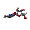

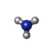

| #2: Chemical | ChemComp-ZN /  Mass: 65.409 Da / Num. of mol.: 4 / Source method: obtained synthetically / Formula: Zn Mass: 65.409 Da / Num. of mol.: 4 / Source method: obtained synthetically / Formula: Zn#3: Chemical | ChemComp-SO4 / |  Mass: 96.063 Da / Num. of mol.: 1 / Source method: obtained synthetically / Formula: SO4 Mass: 96.063 Da / Num. of mol.: 1 / Source method: obtained synthetically / Formula: SO4#4: Chemical |  Mass: 243.217 Da / Num. of mol.: 3 / Source method: obtained synthetically / Formula: C9H13N3O5 Mass: 243.217 Da / Num. of mol.: 3 / Source method: obtained synthetically / Formula: C9H13N3O5#5: Chemical | ChemComp-URI / |  Mass: 244.201 Da / Num. of mol.: 1 / Source method: obtained synthetically / Formula: C9H12N2O6 Mass: 244.201 Da / Num. of mol.: 1 / Source method: obtained synthetically / Formula: C9H12N2O6#6: Chemical | ChemComp-NH3 / |  Mass: 17.031 Da / Num. of mol.: 1 / Source method: obtained synthetically / Formula: NH3 Mass: 17.031 Da / Num. of mol.: 1 / Source method: obtained synthetically / Formula: NH3#7: Water | ChemComp-HOH / | Mass: 18.015 Da / Num. of mol.: 358 / Source method: isolated from a natural source / Formula: H2O |
|---|
-Experimental details
-Experiment
| Experiment | Method: X-RAY DIFFRACTION / Number of used crystals: 1 |
|---|
- Sample preparation
| Crystal | Density Matthews: 2.67 Å3/Da / Density % sol: 53.95 % |
|---|---|
| Crystal grow | Temperature: 298 K / Method: vapor diffusion, hanging drop / pH: 4 Details: 0.9M AMMONIUM SULPHATE, 0.1M CITRATE, pH 4.0, VAPOR DIFFUSION, HANGING DROP, temperature 298.0K |
-Data collection
| Diffraction | Mean temperature: 100 K |
|---|---|
| Diffraction source | Source: ROTATING ANODE / Type: RIGAKU RU300 / Wavelength: 1.5418 Å |
| Detector | Type: RIGAKU RAXIS IV / Detector: IMAGE PLATE / Date: Mar 28, 2004 |
| Radiation | Monochromator: GRAPHITE / Protocol: SINGLE WAVELENGTH / Monochromatic (M) / Laue (L): M / Scattering type: x-ray |
| Radiation wavelength | Wavelength: 1.5418 Å / Relative weight: 1 |
| Reflection | Resolution: 2.07→33.59 Å / Num. all: 42537 / Num. obs: 42537 / % possible obs: 100 % / Observed criterion σ(F): 0 / Observed criterion σ(I): 0 / Redundancy: 4.6 % / Biso Wilson estimate: 17 Å2 / Rmerge(I) obs: 0.079 / Χ2: 0.88 / Net I/σ(I): 11.4 / Scaling rejects: 1478 |
| Reflection shell | Resolution: 2.07→2.14 Å / % possible obs: 99.9 % / Redundancy: 4.53 % / Rmerge(I) obs: 0.345 / Mean I/σ(I) obs: 3.9 / Num. measured all: 19190 / Num. unique all: 4184 / Num. unique obs: 4184 / Χ2: 1.05 / % possible all: 99.9 |
- Processing
| Software |
| ||||||||||||||||||||||||||||||||||||
|---|---|---|---|---|---|---|---|---|---|---|---|---|---|---|---|---|---|---|---|---|---|---|---|---|---|---|---|---|---|---|---|---|---|---|---|---|---|
| Refinement | Method to determine structure: MOLECULAR REPLACEMENT Starting model: PDB ENTRY 2FR5 Resolution: 2.07→33.59 Å / Rfactor Rfree error: 0.005 / Data cutoff high absF: 90351.289 / Data cutoff low absF: 0 / Cross valid method: THROUGHOUT / σ(F): 0 / Stereochemistry target values: Engh & Huber Details: Due to excessive electron density, NH1 of ARG 68 was refined with unit occupancy for both alternate conformations in the four subunits. Disordered atoms were refined with zero occupancy.
| ||||||||||||||||||||||||||||||||||||
| Solvent computation | Solvent model: FLAT MODEL / Bsol: 69.423 Å2 / ksol: 0.374 e/Å3 | ||||||||||||||||||||||||||||||||||||
| Displacement parameters | Biso mean: 25.6 Å2
| ||||||||||||||||||||||||||||||||||||
| Refine analyze |
| ||||||||||||||||||||||||||||||||||||
| Refinement step | Cycle: LAST / Resolution: 2.07→33.59 Å
| ||||||||||||||||||||||||||||||||||||
| Refine LS restraints |
| ||||||||||||||||||||||||||||||||||||
| LS refinement shell | Resolution: 2.07→2.2 Å / Rfactor Rfree error: 0.015 / Total num. of bins used: 6
| ||||||||||||||||||||||||||||||||||||
| Xplor file |
|
