 Movie
Movie Controller

[English] 日本語
 Yorodumi
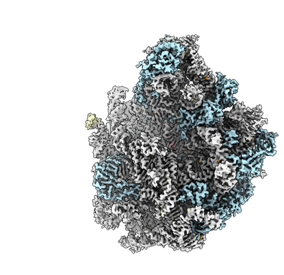
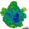
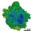
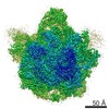
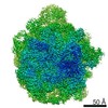
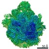
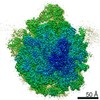
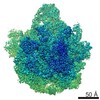
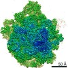
- EMDB-4638: Structure of a bacterial 50S ribosomal subunit in complex with th... -
+ Open data

- Basic information
| Entry | Database: EMDB / ID: EMD-4638 | |||||||||
|---|---|---|---|---|---|---|---|---|---|---|
| Title | Structure of a bacterial 50S ribosomal subunit in complex with the novel quinoxolidinone antibiotic cadazolid | |||||||||
 Map data Map data | ||||||||||
 Sample Sample |
| |||||||||
 Keywords Keywords | ribosome / cadazolid / quinoxolidinone / 50S / ANTIBIOTIC | |||||||||
| Function / homology |  Function and homology information Function and homology informationtranscriptional attenuation / endoribonuclease inhibitor activity / RNA-binding transcription regulator activity / positive regulation of ribosome biogenesis / negative regulation of cytoplasmic translation / DnaA-L2 complex / translation repressor activity / negative regulation of DNA-templated DNA replication initiation / ribosome assembly / mRNA regulatory element binding translation repressor activity ...transcriptional attenuation / endoribonuclease inhibitor activity / RNA-binding transcription regulator activity / positive regulation of ribosome biogenesis / negative regulation of cytoplasmic translation / DnaA-L2 complex / translation repressor activity / negative regulation of DNA-templated DNA replication initiation / ribosome assembly / mRNA regulatory element binding translation repressor activity / assembly of large subunit precursor of preribosome / cytosolic ribosome assembly / regulation of cell growth / response to reactive oxygen species / DNA-templated transcription termination / response to radiation / ribosomal large subunit assembly / mRNA 5'-UTR binding / large ribosomal subunit / ribosome binding / transferase activity / 5S rRNA binding / large ribosomal subunit rRNA binding / cytosolic large ribosomal subunit / tRNA binding / cytoplasmic translation / rRNA binding / negative regulation of translation / ribosome / structural constituent of ribosome / translation / response to antibiotic / negative regulation of DNA-templated transcription / mRNA binding / DNA binding / RNA binding / zinc ion binding / cytoplasm / cytosol Similarity search - Function | |||||||||
| Biological species |  | |||||||||
| Method | single particle reconstruction / cryo EM / Resolution: 3.0 Å | |||||||||
 Authors Authors | Scaiola A / Leibundgut M | |||||||||
 Citation Citation | Journal: Sci Rep / Year: 2019 Title: Structural basis of translation inhibition by cadazolid, a novel quinoxolidinone antibiotic. Authors: Alain Scaiola / Marc Leibundgut / Daniel Boehringer / Patrick Caspers / Daniel Bur / Hans H Locher / Georg Rueedi / Daniel Ritz /  Abstract: Oxazolidinones are synthetic antibiotics used for treatment of infections caused by Gram-positive bacteria. They target the bacterial protein synthesis machinery by binding to the peptidyl ...Oxazolidinones are synthetic antibiotics used for treatment of infections caused by Gram-positive bacteria. They target the bacterial protein synthesis machinery by binding to the peptidyl transferase centre (PTC) of the ribosome and interfering with the peptidyl transferase reaction. Cadazolid is the first member of quinoxolidinone antibiotics, which are characterized by combining the pharmacophores of oxazolidinones and fluoroquinolones, and it is evaluated for treatment of Clostridium difficile gastrointestinal infections that frequently occur in hospitalized patients. In vitro protein synthesis inhibition by cadazolid was shown in Escherichia coli and Staphylococcus aureus, including an isolate resistant against linezolid, the prototypical oxazolidinone antibiotic. To better understand the mechanism of inhibition, we determined a 3.0 Å cryo-electron microscopy structure of cadazolid bound to the E. coli ribosome in complex with mRNA and initiator tRNA. Here we show that cadazolid binds with its oxazolidinone moiety in a binding pocket in close vicinity of the PTC as observed previously for linezolid, and that it extends its unique fluoroquinolone moiety towards the A-site of the PTC. In this position, the drug inhibits protein synthesis by interfering with the binding of tRNA to the A-site, suggesting that its chemical features also can enable the inhibition of linezolid-resistant strains. | |||||||||
| History |
|
- Structure visualization
| Movie |
Movie viewer |
|---|---|
| Structure viewer | EM map: SurfViewMolmilJmol/JSmol |
| Supplemental images |
- Downloads & links
-EMDB archive
| Map data | emd_4638.map.gz | 18 MB | EMDB map data format | |
|---|---|---|---|---|
| Header (meta data) | emd-4638-v30.xmlemd-4638.xml | 46.4 KB 46.4 KB | Display Display | EMDB header |
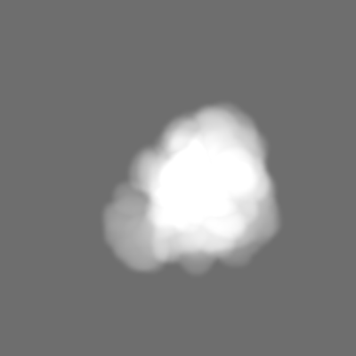
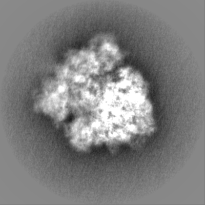
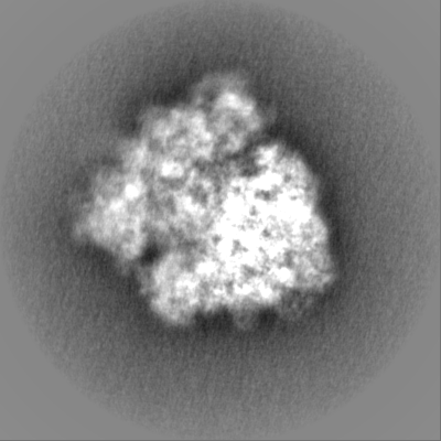
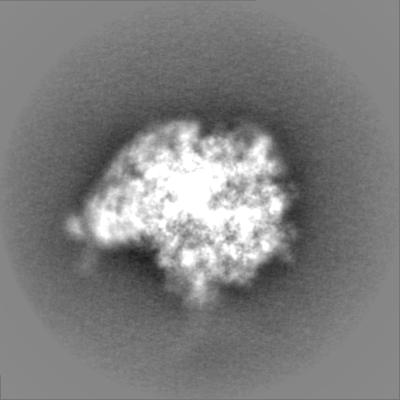
| Images |  emd_4638.png emd_4638.png | 152.4 KB | ||
| Masks | emd_4638_msk_1.map | 244.1 MB | Mask map | |
| Filedesc metadata | emd-4638.cif.gz | 10 KB | ||
| Others | emd_4638_half_map_1.map.gzemd_4638_half_map_2.map.gz | 193 MB 193.2 MB | ||
| Archive directory |  http://ftp.pdbj.org/pub/emdb/structures/EMD-4638ftp://ftp.pdbj.org/pub/emdb/structures/EMD-4638 http://ftp.pdbj.org/pub/emdb/structures/EMD-4638ftp://ftp.pdbj.org/pub/emdb/structures/EMD-4638 | HTTPS FTP |
-Validation report
| Summary document | emd_4638_validation.pdf.gz | 412.8 KB | Display | EMDB validaton report |
|---|---|---|---|---|
| Full document | emd_4638_full_validation.pdf.gz | 412 KB | Display | |
| Data in XML | emd_4638_validation.xml.gz | 494 B | Display | |
| Arichive directory | https://ftp.pdbj.org/pub/emdb/validation_reports/EMD-4638ftp://ftp.pdbj.org/pub/emdb/validation_reports/EMD-4638 | HTTPS FTP |
-Related structure data
| Related structure data |  6qulMC  4639C  4641C M: atomic model generated by this map C: citing same article ( |
|---|---|
| Similar structure data |


-Links
| EMDB pages | EMDB (EBI/PDBe) / EMDataResource |
|---|---|
| Related items in Molecule of the Month |


-Map
| File | Download / File: emd_4638.map.gz / Format: CCP4 / Size: 244.1 MB / Type: IMAGE STORED AS FLOATING POINT NUMBER (4 BYTES) | ||||||||||||||||||||||||||||||||||||||||||||||||||||||||||||||||||||
|---|---|---|---|---|---|---|---|---|---|---|---|---|---|---|---|---|---|---|---|---|---|---|---|---|---|---|---|---|---|---|---|---|---|---|---|---|---|---|---|---|---|---|---|---|---|---|---|---|---|---|---|---|---|---|---|---|---|---|---|---|---|---|---|---|---|---|---|---|---|
| Voxel size | X=Y=Z: 1.079 Å | ||||||||||||||||||||||||||||||||||||||||||||||||||||||||||||||||||||
| Density |
| ||||||||||||||||||||||||||||||||||||||||||||||||||||||||||||||||||||
| Symmetry | Space group: 1 | ||||||||||||||||||||||||||||||||||||||||||||||||||||||||||||||||||||
| Details | EMDB XML:
CCP4 map header:
| ||||||||||||||||||||||||||||||||||||||||||||||||||||||||||||||||||||
-Supplemental data
-Mask #1
| File | emd_4638_msk_1.map | ||||||||||||
|---|---|---|---|---|---|---|---|---|---|---|---|---|---|
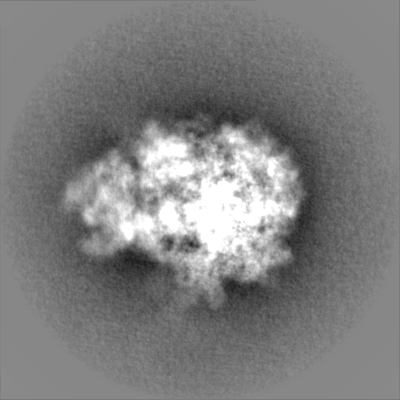
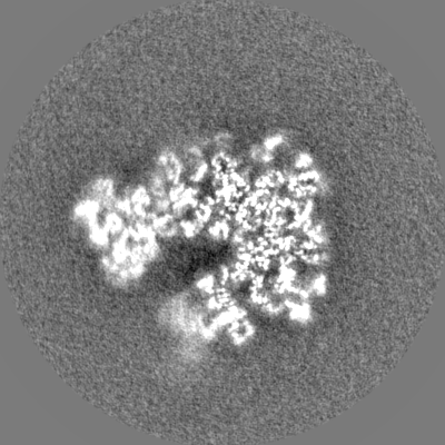
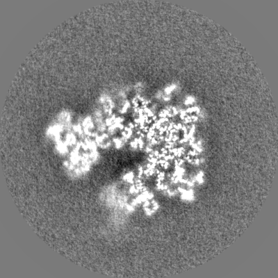
| Projections & Slices |
| ||||||||||||
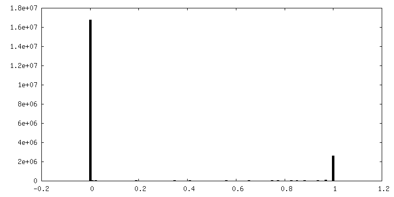
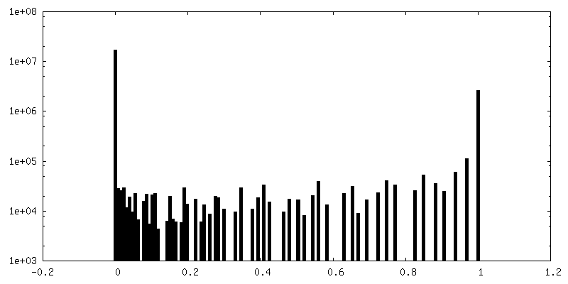
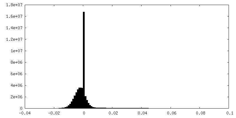
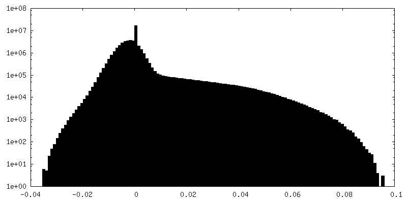
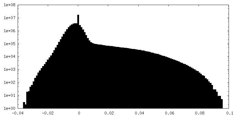
| Density Histograms |
 Z
Z Y
Y X
X


-Half map: #1
| File | emd_4638_half_map_1.map | ||||||||||||
|---|---|---|---|---|---|---|---|---|---|---|---|---|---|
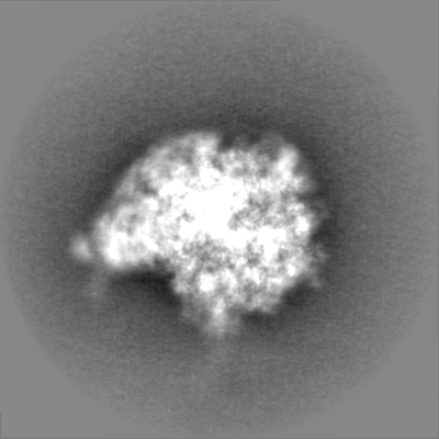
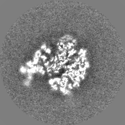
| Projections & Slices |
| ||||||||||||
| Density Histograms |

-Half map: #2
| File | emd_4638_half_map_2.map | ||||||||||||
|---|---|---|---|---|---|---|---|---|---|---|---|---|---|
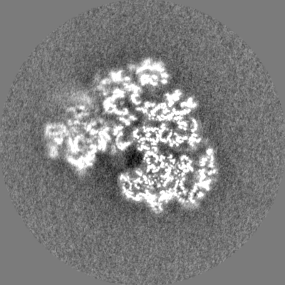
| Projections & Slices |
| ||||||||||||
| Density Histograms |


- Sample components
+Entire : Structure of a bacterial 50S ribosomal subunit in complex with th...
+Supramolecule #1: Structure of a bacterial 50S ribosomal subunit in complex with th...
+Macromolecule #1: P-site fMet-tRNA(fMet)
+Macromolecule #2: 23S rRNA
+Macromolecule #3: 5S rRNA
+Macromolecule #4: 50S ribosomal protein L2
+Macromolecule #5: 50S ribosomal protein L3
+Macromolecule #6: 50S ribosomal protein L4
+Macromolecule #7: 50S ribosomal protein L5
+Macromolecule #8: 50S ribosomal protein L6
+Macromolecule #9: 50S ribosomal protein L9
+Macromolecule #10: 50S ribosomal protein L13
+Macromolecule #11: 50S ribosomal protein L14
+Macromolecule #12: 50S ribosomal protein L15
+Macromolecule #13: 50S ribosomal protein L16
+Macromolecule #14: 50S ribosomal protein L17
+Macromolecule #15: 50S ribosomal protein L18
+Macromolecule #16: 50S ribosomal protein L19
+Macromolecule #17: 50S ribosomal protein L20
+Macromolecule #18: 50S ribosomal protein L21
+Macromolecule #19: 50S ribosomal protein L22
+Macromolecule #20: 50S ribosomal protein L23
+Macromolecule #21: 50S ribosomal protein L24
+Macromolecule #22: 50S ribosomal protein L25
+Macromolecule #23: 50S ribosomal protein L27
+Macromolecule #24: 50S ribosomal protein L28
+Macromolecule #25: 50S ribosomal protein L29
+Macromolecule #26: 50S ribosomal protein L30
+Macromolecule #27: 50S ribosomal protein L32
+Macromolecule #28: 50S ribosomal protein L33
+Macromolecule #29: 50S ribosomal protein L34
+Macromolecule #30: 50S ribosomal protein L35
+Macromolecule #31: 50S ribosomal protein L36
+Macromolecule #32: N-FORMYLMETHIONINE
+Macromolecule #33: MAGNESIUM ION
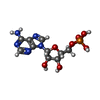
+Macromolecule #34: ADENOSINE-5'-MONOPHOSPHATE
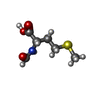
+Macromolecule #35: cadazolid

+Macromolecule #36: ZINC ION
-Experimental details
-Structure determination
| Method | cryo EM |
|---|---|
 Processing Processing | single particle reconstruction |
| Aggregation state | particle |
-Sample preparation
| Buffer | pH: 7.4 |
|---|---|
| Grid | Model: Quantifoil R2/2 / Material: COPPER |
| Vitrification | Cryogen name: ETHANE-PROPANE / Chamber humidity: 100 % / Chamber temperature: 277 K / Instrument: FEI VITROBOT MARK IV |
- Electron microscopy
| Microscope | FEI TITAN KRIOS |
|---|---|
| Image recording | Film or detector model: FEI FALCON II (4k x 4k) / Detector mode: INTEGRATING / Average electron dose: 1.14 e/Å2 |
| Electron beam | Acceleration voltage: 300 kV / Electron source:  FIELD EMISSION GUN FIELD EMISSION GUN |
| Electron optics | Illumination mode: FLOOD BEAM / Imaging mode: BRIGHT FIELD |
| Experimental equipment |  Model: Titan Krios / Image courtesy: FEI Company |
-Image processing
| Startup model | Type of model: PDB ENTRY PDB model - PDB ID: |
|---|---|
| Final reconstruction | Applied symmetry - Point group: C1 (asymmetric) / Resolution.type: BY AUTHOR / Resolution: 3.0 Å / Resolution method: FSC 0.143 CUT-OFF / Software - Name: RELION (ver. 3.0) / Number images used: 59148 |
| Initial angle assignment | Type: MAXIMUM LIKELIHOOD / Software - Name: RELION (ver. 2.0) |
| Final angle assignment | Type: MAXIMUM LIKELIHOOD / Software - Name: RELION (ver. 3.0) |

