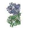
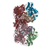
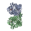
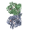
[English] 日本語
Yorodumi
- PDB-3etr: Crystal structure of xanthine oxidase in complex with lumazine -

+
Open data


ID or keywords:

Loading...

-
Basic information

Entry
Database: PDB / ID: 3etr
TitleCrystal structure of xanthine oxidase in complex with lumazine
Components(Xanthine dehydrogenase/oxidase) x 3
KeywordsOXIDOREDUCTASE / protein-ligand complex / enzyme catalysis / substrate orientation / FAD / Flavoprotein / Iron / Iron-sulfur / Metal-binding / Molybdenum / NAD / Peroxisome
Function / homology
Function and homology information


xanthine dehydrogenase complex / xanthine dehydrogenase / xanthine oxidase / xanthine oxidase activity / xanthine catabolic process / xanthine dehydrogenase activity / molybdenum ion binding / molybdopterin cofactor binding / FAD binding / 2 iron, 2 sulfur cluster binding ...xanthine dehydrogenase complex / xanthine dehydrogenase / xanthine oxidase / xanthine oxidase activity / xanthine catabolic process / xanthine dehydrogenase activity / molybdenum ion binding / molybdopterin cofactor binding / FAD binding / 2 iron, 2 sulfur cluster binding / peroxisome / flavin adenine dinucleotide binding / iron ion binding / protein homodimerization activity / extracellular space
Similarity search - Function
Xanthine dehydrogenase, small subunit / Oxidoreductase, molybdopterin binding site / Eukaryotic molybdopterin oxidoreductases signature. / Aldehyde oxidase/xanthine dehydrogenase, a/b hammerhead / CO dehydrogenase flavoprotein, C-terminal domain / CO dehydrogenase flavoprotein C-terminal domain / Aldehyde Oxidoreductase; domain 4 / Aldehyde oxidase/xanthine dehydrogenase, molybdopterin binding domain / [2Fe-2S]-binding domain / Molybdopterin dehydrogenase, FAD-binding ...Xanthine dehydrogenase, small subunit / Oxidoreductase, molybdopterin binding site / Eukaryotic molybdopterin oxidoreductases signature. / Aldehyde oxidase/xanthine dehydrogenase, a/b hammerhead / CO dehydrogenase flavoprotein, C-terminal domain / CO dehydrogenase flavoprotein C-terminal domain / Aldehyde Oxidoreductase; domain 4 / Aldehyde oxidase/xanthine dehydrogenase, molybdopterin binding domain / [2Fe-2S]-binding domain / Molybdopterin dehydrogenase, FAD-binding / CO dehydrogenase flavoprotein, C-terminal / CO dehydrogenase flavoprotein, C-terminal domain superfamily / FAD binding domain in molybdopterin dehydrogenase / CO dehydrogenase flavoprotein C-terminal domain / Aldehyde oxidase/xanthine dehydrogenase / Aldehyde oxidase/xanthine dehydrogenase, second molybdopterin binding domain / Molybdopterin cofactor-binding domain / Aldehyde oxidase and xanthine dehydrogenase, a/b hammerhead domain / Aldehyde oxidase/xanthine dehydrogenase, a/b hammerhead / [2Fe-2S]-binding / Aldehyde oxidase/xanthine dehydrogenase, first molybdopterin binding domain / Aldehyde oxidase/xanthine dehydrogenase, a/b hammerhead superfamily / [2Fe-2S]-binding domain superfamily / Aldehyde oxidase/xanthine dehydrogenase, molybdopterin binding domain superfamily / [2Fe-2S] binding domain / Molybdopterin cofactor-binding domain / Aldehyde oxidase and xanthine dehydrogenase, a/b hammerhead domain / Uridine Diphospho-n-acetylenolpyruvylglucosamine Reductase, domain 2 / Uridine Diphospho-n-acetylenolpyruvylglucosamine Reductase; domain 2 / Uridine Diphospho-n-acetylenolpyruvylglucosamine Reductase; domain 3 - #10 / Uridine Diphospho-n-acetylenolpyruvylglucosamine Reductase; domain 3 / FAD-binding, type PCMH, subdomain 1 / Aldehyde Oxidoreductase; domain 3 / 2Fe-2S ferredoxin, iron-sulphur binding site / 2Fe-2S ferredoxin-type iron-sulfur binding region signature. / FAD-binding domain, PCMH-type / PCMH-type FAD-binding domain profile. / Beta-grasp domain / FAD-binding, type PCMH, subdomain 2 / 2Fe-2S iron-sulfur cluster binding domain / FAD-binding, type PCMH-like superfamily / Beta-grasp domain superfamily / Enolase-like; domain 1 / 2Fe-2S ferredoxin-type iron-sulfur binding domain profile. / 2Fe-2S ferredoxin-type iron-sulfur binding domain / 2Fe-2S ferredoxin-like superfamily / Ubiquitin-like (UB roll) / DNA polymerase; domain 1 / Roll / Alpha-Beta Complex / 2-Layer Sandwich / Orthogonal Bundle / Mainly Alpha / Alpha Beta
Similarity search - Domain/homology
FLAVIN-ADENINE DINUCLEOTIDE / FE2/S2 (INORGANIC) CLUSTER / pteridine-2,4(1H,3H)-dione / DIOXOTHIOMOLYBDENUM(VI) ION / Chem-MTE / Xanthine dehydrogenase/oxidase
Similarity search - Component
Biological speciesBos taurus (cattle)
MethodX-RAY DIFFRACTION / SYNCHROTRON / MOLECULAR REPLACEMENT / molecular replacement / Resolution: 2.2 Å
AuthorsPauff, J.M. / Cao, H. / Hille, R.
CitationJournal: J.Biol.Chem. / Year: 2009
Title: Substrate Orientation and Catalysis at the Molybdenum Site in Xanthine Oxidase: CRYSTAL STRUCTURES IN COMPLEX WITH XANTHINE AND LUMAZINE.
Authors: Pauff, J.M. / Cao, H. / Hille, R.
History
DepositionOct 8, 2008Deposition site: RCSB / Processing site: RCSB
Revision 1.0Jan 27, 2009Provider: repository / Type: Initial release
Revision 1.1Jul 13, 2011Group: Version format compliance
Revision 1.2Sep 10, 2014Group: Database references
Revision 1.3Oct 25, 2017Group: Refinement description / Category: software / Item: _software.name
Revision 1.4Sep 6, 2023Group: Data collection / Database references ...Data collection / Database references / Derived calculations / Refinement description / Structure summary
Category: chem_comp / chem_comp_atom ...chem_comp / chem_comp_atom / chem_comp_bond / database_2 / pdbx_initial_refinement_model / pdbx_struct_conn_angle / struct_conn / struct_site
Item: _chem_comp.pdbx_synonyms / _database_2.pdbx_DOI ..._chem_comp.pdbx_synonyms / _database_2.pdbx_DOI / _database_2.pdbx_database_accession / _pdbx_struct_conn_angle.ptnr1_auth_asym_id / _pdbx_struct_conn_angle.ptnr1_auth_comp_id / _pdbx_struct_conn_angle.ptnr1_auth_seq_id / _pdbx_struct_conn_angle.ptnr1_label_asym_id / _pdbx_struct_conn_angle.ptnr1_label_atom_id / _pdbx_struct_conn_angle.ptnr1_label_comp_id / _pdbx_struct_conn_angle.ptnr1_label_seq_id / _pdbx_struct_conn_angle.ptnr2_auth_asym_id / _pdbx_struct_conn_angle.ptnr2_auth_comp_id / _pdbx_struct_conn_angle.ptnr2_auth_seq_id / _pdbx_struct_conn_angle.ptnr2_label_asym_id / _pdbx_struct_conn_angle.ptnr2_label_atom_id / _pdbx_struct_conn_angle.ptnr2_label_comp_id / _pdbx_struct_conn_angle.ptnr3_auth_asym_id / _pdbx_struct_conn_angle.ptnr3_auth_comp_id / _pdbx_struct_conn_angle.ptnr3_auth_seq_id / _pdbx_struct_conn_angle.ptnr3_label_asym_id / _pdbx_struct_conn_angle.ptnr3_label_atom_id / _pdbx_struct_conn_angle.ptnr3_label_comp_id / _pdbx_struct_conn_angle.ptnr3_label_seq_id / _pdbx_struct_conn_angle.value / _struct_conn.pdbx_dist_value / _struct_conn.ptnr1_auth_asym_id / _struct_conn.ptnr1_auth_comp_id / _struct_conn.ptnr1_auth_seq_id / _struct_conn.ptnr1_label_asym_id / _struct_conn.ptnr1_label_atom_id / _struct_conn.ptnr1_label_comp_id / _struct_conn.ptnr1_label_seq_id / _struct_conn.ptnr2_auth_asym_id / _struct_conn.ptnr2_auth_comp_id / _struct_conn.ptnr2_auth_seq_id / _struct_conn.ptnr2_label_asym_id / _struct_conn.ptnr2_label_atom_id / _struct_conn.ptnr2_label_comp_id / _struct_site.pdbx_auth_asym_id / _struct_site.pdbx_auth_comp_id / _struct_site.pdbx_auth_seq_id

-
Structure visualization

Structure viewerMolecule:
MolmilJmol/JSmol

Downloads & links

-
Assembly

Deposited unit
A: Xanthine dehydrogenase/oxidase
B: Xanthine dehydrogenase/oxidase
C: Xanthine dehydrogenase/oxidase
L: Xanthine dehydrogenase/oxidase
M: Xanthine dehydrogenase/oxidase
N: Xanthine dehydrogenase/oxidase
hetero molecules


Theoretical massNumber of molelcules
Total (without water)273,74319
Polymers269,9876
Non-polymers3,75513
Water22,1941232
1


  • Idetical with deposited unit
  • defined by author&software
TypeNameSymmetry operationNumber
identity operation1_555x,y,z1
Buried area24080 Å2
ΔGint-83.2 kcal/mol
Surface area81930 Å2
MethodPISA
2
A: Xanthine dehydrogenase/oxidase
B: Xanthine dehydrogenase/oxidase
C: Xanthine dehydrogenase/oxidase
hetero molecules


Theoretical massNumber of molelcules
Total (without water)136,89110
Polymers134,9943
Non-polymers1,8987
Water543
TypeNameSymmetry operationNumber
identity operation1_555x,y,z1
Buried area8930 Å2
ΔGint-37.4 kcal/mol
Surface area43820 Å2
MethodPISA
3
L: Xanthine dehydrogenase/oxidase
M: Xanthine dehydrogenase/oxidase
N: Xanthine dehydrogenase/oxidase
hetero molecules


Theoretical massNumber of molelcules
Total (without water)136,8519
Polymers134,9943
Non-polymers1,8586
Water543
TypeNameSymmetry operationNumber
identity operation1_555x,y,z1
Buried area9210 Å2
ΔGint-38.6 kcal/mol
Surface area44050 Å2
MethodPISA
Unit cell
Length a, b, c (Å)133.190, 73.491, 146.505
Angle α, β, γ (deg.)90.000, 98.680, 90.000
Int Tables number4
Space group name H-MP1211
DetailsBIOLOGICAL UNIT IS THE SAME AS ASYMMETRIC UNIT.

-
Components

-
Protein , 3 types, 6 molecules ALBMCN

#1: Protein Xanthine dehydrogenase/oxidase / [Includes: Xanthine dehydrogenase (EC 1.17.1.4) (XD) / and Xanthine oxidase (EC 1.17.3.2) (XO) ...[Includes: Xanthine dehydrogenase (EC 1.17.1.4) (XD) / and Xanthine oxidase (EC 1.17.3.2) (XO) (Xanthine oxidoreductase)]


Mass: 18002.881 Da / Num. of mol.: 2 / Fragment: UNP residues 2-165 / Source method: isolated from a natural source / Details: Animal Milk / Source: (natural) Bos taurus (cattle) / References: UniProt: P80457
#2: Protein Xanthine dehydrogenase/oxidase


Mass: 34024.590 Da / Num. of mol.: 2 / Fragment: UNP residues 224-528 / Source method: isolated from a natural source / Details: Animal Milk / Source: (natural) Bos taurus (cattle) / References: UniProt: P80457
#3: Protein Xanthine dehydrogenase/oxidase


Mass: 82966.203 Da / Num. of mol.: 2 / Fragment: UNP residues 571-1325 / Source method: isolated from a natural source / Details: Animal Milk / Source: (natural) Bos taurus (cattle) / References: UniProt: P80457

-
Non-polymers , 7 types, 1245 molecules

#4: Chemical
ChemComp-FES / FE2/S2 (INORGANIC) CLUSTER / Iron–sulfur cluster


Mass: 175.820 Da / Num. of mol.: 4 / Source method: obtained synthetically / Formula: Fe2S2
#5: Chemical ChemComp-FAD / FLAVIN-ADENINE DINUCLEOTIDE / Flavin adenine dinucleotide


Mass: 785.550 Da / Num. of mol.: 2 / Source method: obtained synthetically / Formula: C27H33N9O15P2 / Comment: FAD*YM
#6: Chemical ChemComp-MTE / PHOSPHONIC ACIDMONO-(2-AMINO-5,6-DIMERCAPTO-4-OXO-3,7,8A,9,10,10A-HEXAHYDRO-4H-8-OXA-1,3,9,10-TETRAAZA-ANTHRACEN-7-YLMETHYL)ESTER


Mass: 395.352 Da / Num. of mol.: 2 / Source method: obtained synthetically / Formula: C10H14N5O6PS2
#7: Chemical ChemComp-MOS / DIOXOTHIOMOLYBDENUM(VI) ION


Mass: 161.012 Da / Num. of mol.: 2 / Source method: obtained synthetically / Formula: HMoO2S
#8: Chemical ChemComp-LUZ / pteridine-2,4(1H,3H)-dione / Lumazine / 2,4-dihydroxypteridine / 2,4-pteridinediol


Mass: 164.122 Da / Num. of mol.: 2 / Source method: obtained synthetically / Formula: C6H4N4O2
#9: Chemical ChemComp-CA / CALCIUM ION


Mass: 40.078 Da / Num. of mol.: 1 / Source method: obtained synthetically / Formula: Ca
#10: Water ChemComp-HOH / water / Water


Mass: 18.015 Da / Num. of mol.: 1232 / Source method: isolated from a natural source / Formula: H2O

-
Experimental details

-
Experiment

ExperimentMethod: X-RAY DIFFRACTION / Number of used crystals: 1

-
Sample preparation

CrystalDensity Matthews: 2.63 Å3/Da / Density % sol: 53.15 %
Crystal growTemperature: 298 K / Method: batch / pH: 7.2 / Details: PEG 8000, pH 7.2, BATCH, temperature 298K

-
Data collection

DiffractionMean temperature: 100 K
Diffraction sourceSource: SYNCHROTRON / Site: APS / Beamline: 31-ID / Wavelength: 0.9793 Å
DetectorType: MARMOSAIC 325 mm CCD / Detector: CCD / Date: Jun 15, 2007 / Details: APS BEAMLINE SGX PHARMACEUTICALS
RadiationMonochromator: Si(111) / Protocol: SINGLE WAVELENGTH / Monochromatic (M) / Laue (L): M / Scattering type: x-ray
Radiation wavelengthWavelength: 0.9793 Å / Relative weight: 1
ReflectionResolution: 2.2→50 Å / Num. obs: 140747 / % possible obs: 98.7 % / Biso Wilson estimate: 13.1 Å2 / Rsym value: 0.197 / Net I/σ(I): 5
Reflection shellResolution: 2.2→2.257 Å / % possible obs: 96.1 % / Mean I/σ(I) obs: 1.7

-
Phasing

PhasingMethod: molecular replacement
Phasing MR
Highest resolutionLowest resolution
Rotation2.11 Å26.42 Å
Translation2.11 Å26.42 Å

-
Processing

Software
NameVersionClassificationNB
MOLREPphasing
REFMACrefinement
PDB_EXTRACT3.006data extraction
MAR345CCDdata collection
HKL-2000data reduction
HKL-2000data scaling
RefinementMethod to determine structure: MOLECULAR REPLACEMENT
Starting model: PDB entry 1FIQ
Resolution: 2.2→50 Å / Cor.coef. Fo:Fc: 0.942 / Cor.coef. Fo:Fc free: 0.893 / WRfactor Rfree: 0.283 / WRfactor Rwork: 0.209 / Occupancy max: 1 / Occupancy min: 1 / FOM work R set: 0.803 / SU B: 6.927 / SU ML: 0.176 / SU R Cruickshank DPI: 0.276 / SU Rfree: 0.234 / Cross valid method: THROUGHOUT / σ(F): 0 / ESU R: 0.276 / ESU R Free: 0.234 / Stereochemistry target values: MAXIMUM LIKELIHOOD / Details: HYDROGENS HAVE BEEN ADDED IN THE RIDING POSITIONS
RfactorNum. reflection% reflectionSelection details
Rfree0.267 7052 5 %RANDOM
Rwork0.197 ---
obs0.201 140747 98.71 %-
Solvent computationIon probe radii: 0.8 Å / Shrinkage radii: 0.8 Å / VDW probe radii: 1.2 Å / Solvent model: MASK
Displacement parametersBiso max: 84.91 Å2 / Biso mean: 29.464 Å2 / Biso min: 2 Å2
Baniso -1Baniso -2Baniso -3
1--2.54 Å20 Å21.16 Å2
2--0.08 Å20 Å2
3---2.81 Å2
Refinement stepCycle: LAST / Resolution: 2.2→50 Å
ProteinNucleic acidLigandSolventTotal
Num. atoms18872 0 203 1232 20307
Refine LS restraints
Refine-IDTypeDev idealDev ideal targetNumber
X-RAY DIFFRACTIONr_bond_refined_d0.020.02219485
X-RAY DIFFRACTIONr_angle_refined_deg1.961.97426377
X-RAY DIFFRACTIONr_dihedral_angle_1_deg7.96752432
X-RAY DIFFRACTIONr_dihedral_angle_2_deg39.12123.791815
X-RAY DIFFRACTIONr_dihedral_angle_3_deg18.192153337
X-RAY DIFFRACTIONr_dihedral_angle_4_deg19.31815118
X-RAY DIFFRACTIONr_chiral_restr0.1290.22953
X-RAY DIFFRACTIONr_gen_planes_refined0.0080.0214540
X-RAY DIFFRACTIONr_nbd_refined0.2380.29387
X-RAY DIFFRACTIONr_nbtor_refined0.3080.213095
X-RAY DIFFRACTIONr_xyhbond_nbd_refined0.2050.21322
X-RAY DIFFRACTIONr_metal_ion_refined0.6110.21
X-RAY DIFFRACTIONr_symmetry_vdw_refined0.2630.228
X-RAY DIFFRACTIONr_symmetry_hbond_refined0.290.23
X-RAY DIFFRACTIONr_mcbond_it1.0011.512489
X-RAY DIFFRACTIONr_mcangle_it1.609219520
X-RAY DIFFRACTIONr_scbond_it2.66438105
X-RAY DIFFRACTIONr_scangle_it4.0074.56849
LS refinement shellResolution: 2.2→2.257 Å / Total num. of bins used: 20
RfactorNum. reflection% reflection
Rfree0.342 496 -
Rwork0.236 9645 -
all-10141 -
obs--96.11 %

+
About Yorodumi

-
News

-
Feb 9, 2022. New format data for meta-information of EMDB entries

New format data for meta-information of EMDB entries

  • Version 3 of the EMDB header file is now the official format.
  • The previous official version 1.9 will be removed from the archive.

Related info.:EMDB header

External links:wwPDB to switch to version 3 of the EMDB data model

-
Aug 12, 2020. Covid-19 info

Covid-19 info

URL: https://pdbj.org/emnavi/covid19.php

New page: Covid-19 featured information page in EM Navigator.

Related info.:Covid-19 info / Mar 5, 2020. Novel coronavirus structure data

+
Mar 5, 2020. Novel coronavirus structure data

Novel coronavirus structure data

Related info.:Yorodumi Speices / Aug 12, 2020. Covid-19 info

External links:COVID-19 featured content - PDBj / Molecule of the Month (242):Coronavirus Proteases

+
Jan 31, 2019. EMDB accession codes are about to change! (news from PDBe EMDB page)

EMDB accession codes are about to change! (news from PDBe EMDB page)

  • The allocation of 4 digits for EMDB accession codes will soon come to an end. Whilst these codes will remain in use, new EMDB accession codes will include an additional digit and will expand incrementally as the available range of codes is exhausted. The current 4-digit format prefixed with “EMD-” (i.e. EMD-XXXX) will advance to a 5-digit format (i.e. EMD-XXXXX), and so on. It is currently estimated that the 4-digit codes will be depleted around Spring 2019, at which point the 5-digit format will come into force.
  • The EM Navigator/Yorodumi systems omit the EMD- prefix.

Related info.:Q: What is EMD? / ID/Accession-code notation in Yorodumi/EM Navigator

External links:EMDB Accession Codes are Changing Soon! / Contact to PDBj

+
Jul 12, 2017. Major update of PDB

Major update of PDB

  • wwPDB released updated PDB data conforming to the new PDBx/mmCIF dictionary.
  • This is a major update changing the version number from 4 to 5, and with Remediation, in which all the entries are updated.
  • In this update, many items about electron microscopy experimental information are reorganized (e.g. em_software).
  • Now, EM Navigator and Yorodumi are based on the updated data.

External links:wwPDB Remediation / Enriched Model Files Conforming to OneDep Data Standards Now Available in the PDB FTP Archive

-
Yorodumi

Thousand views of thousand structures

  • Yorodumi is a browser for structure data from EMDB, PDB, SASBDB, etc.
  • This page is also the successor to EM Navigator detail page, and also detail information page/front-end page for Omokage search.
  • The word "yorodu" (or yorozu) is an old Japanese word meaning "ten thousand". "mi" (miru) is to see.

Related info.:EMDB / PDB / SASBDB / Comparison of 3 databanks / Yorodumi Search / Aug 31, 2016. New EM Navigator & Yorodumi / Yorodumi Papers / Jmol/JSmol / Function and homology information / Changes in new EM Navigator and Yorodumi

Read more