 Movie
Movie Controller

[English] 日本語
 Yorodumi
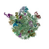
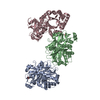
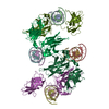
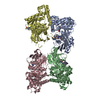
- PDB-2vrh: Structure of the E. coli trigger factor bound to a translating ri... -
+ Open data

- Basic information
| Entry | Database: PDB / ID: 2vrh | ||||||
|---|---|---|---|---|---|---|---|
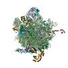
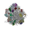
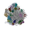
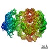
| Title | Structure of the E. coli trigger factor bound to a translating ribosome | ||||||
 Components Components |
| ||||||
 Keywords Keywords | RIBOSOME / TRIGGER FACTOR / RIBOSOMAL PROTEIN / RIBONUCLEOPROTEIN / CO-TRANSLATIONAL PROTEIN FOLDING / ROTAMASE / CHAPERONE / ISOMERASE / CELL CYCLE / RNA-BINDING / RRNA-BINDING / CELL DIVISION / RIBOSOME-NASCENT CHAIN COMPLEX | ||||||
| Function / homology |  Function and homology information Function and homology information'de novo' cotranslational protein folding / stress response to copper ion / protein unfolding / protein folding chaperone / peptidylprolyl isomerase / peptidyl-prolyl cis-trans isomerase activity / protein transport / response to heat / protein folding / ribosome binding ...'de novo' cotranslational protein folding / stress response to copper ion / protein unfolding / protein folding chaperone / peptidylprolyl isomerase / peptidyl-prolyl cis-trans isomerase activity / protein transport / response to heat / protein folding / ribosome binding / ribosomal large subunit assembly / large ribosomal subunit rRNA binding / cytosolic large ribosomal subunit / cytoplasmic translation / rRNA binding / structural constituent of ribosome / ribosome / translation / ribonucleoprotein complex / cell division / membrane / identical protein binding / cytoplasm / cytosol Similarity search - Function | ||||||
| Biological species |  | ||||||
| Method | ELECTRON MICROSCOPY / single particle reconstruction / cryo EM / Resolution: 19 Å | ||||||
| Model type details | CA ATOMS ONLY, CHAIN A, B, C, D | ||||||
 Authors Authors | Merz, F. / Boehringer, D. / Schaffitzel, C. / Preissler, S. / Hoffmann, A. / Maier, T. / Rutkowska, A. / Lozza, J. / Ban, N. / Bukau, B. / Deuerling, E. | ||||||
 Citation Citation | Journal: EMBO J / Year: 2008 Title: Molecular mechanism and structure of Trigger Factor bound to the translating ribosome. Authors: Frieder Merz / Daniel Boehringer / Christiane Schaffitzel / Steffen Preissler / Anja Hoffmann / Timm Maier / Anna Rutkowska / Jasmin Lozza / Nenad Ban / Bernd Bukau / Elke Deuerling /  Abstract: Ribosome-associated chaperone Trigger Factor (TF) initiates folding of newly synthesized proteins in bacteria. Here, we pinpoint by site-specific crosslinking the sequence of molecular interactions ...Ribosome-associated chaperone Trigger Factor (TF) initiates folding of newly synthesized proteins in bacteria. Here, we pinpoint by site-specific crosslinking the sequence of molecular interactions of Escherichia coli TF and nascent chains during translation. Furthermore, we provide the first full-length structure of TF associated with ribosome-nascent chain complexes by using cryo-electron microscopy. In its active state, TF arches over the ribosomal exit tunnel accepting nascent chains in a protective void. The growing nascent chain initially follows a predefined path through the entire interior of TF in an unfolded conformation, and even after folding into a domain it remains accommodated inside the protective cavity of ribosome-bound TF. The adaptability to accept nascent chains of different length and folding states may explain how TF is able to assist co-translational folding of all kinds of nascent polypeptides during ongoing synthesis. Moreover, we suggest a model of how TF's chaperoning function can be coordinated with the co-translational processing and membrane targeting of nascent polypeptides by other ribosome-associated factors. | ||||||
| History |
|
- Structure visualization
| Movie |
Movie viewer |
|---|---|
| Structure viewer | Molecule: MolmilJmol/JSmol |
- Downloads & links
-Download
| PDBx/mmCIF format | 2vrh.cif.gz | 36.9 KB | Display | PDBx/mmCIF format |
|---|---|---|---|---|
| PDB format | pdb2vrh.ent.gz | 19 KB | Display | PDB format |
| PDBx/mmJSON format | 2vrh.json.gz | Tree view | PDBx/mmJSON format | |
| Others |  Other downloads Other downloads |
-Validation report
| Arichive directory | https://data.pdbj.org/pub/pdb/validation_reports/vr/2vrhftp://data.pdbj.org/pub/pdb/validation_reports/vr/2vrh | HTTPS FTP |
|---|
-Related structure data
| Related structure data | 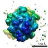 1499MC M: map data used to model this data C: citing same article ( |
|---|---|
| Similar structure data |


-Links
 PDBj
PDBj


- Assembly
| Deposited unit | 
|
|---|---|
| 1 |
|
-Components
| #1: Protein | Mass: 48771.410 Da / Num. of mol.: 1 Source method: isolated from a genetically manipulated source Details: BASED ON PDB 1W26_A. RESIDUES 22-62 WERE REPLACED WITH THE CORRESPONDING RESIDUES OF PDB 1OMS_B Source: (gene. exp.) |
|---|---|
| #2: Protein | Mass: 11222.160 Da / Num. of mol.: 1 / Source method: isolated from a natural source / Details: BASED ON PDB 2AW4_T / Source: (natural) |
| #3: Protein | Mass: 11208.054 Da / Num. of mol.: 1 / Fragment: RESIDUES 2-104 / Source method: isolated from a natural source / Details: BASED ON PDB 2AW4_U / Source: (natural) |
| #4: Protein | Mass: 7286.464 Da / Num. of mol.: 1 / Source method: isolated from a natural source / Details: BASED ON PDB 2AW4_X / Source: (natural) |
| Has protein modification | N |
-Experimental details
-Experiment
| Experiment | Method: ELECTRON MICROSCOPY |
|---|---|
| EM experiment | Aggregation state: PARTICLE / 3D reconstruction method: single particle reconstruction |
- Sample preparation
| Component | Name: STRUCTURE OF THE E. COLI TRIGGER FACTOR BOUND TO A TRANSLATING RIBOSOME Type: RIBOSOME |
|---|---|
| Buffer solution | Name: 50 MM HEPES-KOH PH 7.5, 100 MM KCL, 25 MM MGCL2, 0.5 MG/ML CHLORAMPHENICOL pH: 7.5 Details: 50 MM HEPES-KOH PH 7.5, 100 MM KCL, 25 MM MGCL2, 0.5 MG/ML CHLORAMPHENICOL |
| Specimen | Embedding applied: NO / Shadowing applied: NO / Staining applied: NO / Vitrification applied: YES |
| Specimen support | Details: HOLEY CARBON |
| Vitrification | Cryogen name: ETHANE / Details: LIQUID ETHANE |
- Electron microscopy imaging
| Microscopy | Model: FEI TECNAI 20 |
|---|---|
| Electron gun | Electron source:  FIELD EMISSION GUN / Accelerating voltage: 200 kV / Illumination mode: FLOOD BEAM FIELD EMISSION GUN / Accelerating voltage: 200 kV / Illumination mode: FLOOD BEAM |
| Electron lens | Mode: BRIGHT FIELD / Nominal magnification: 50000 X / Calibrated magnification: 50000 X / Nominal defocus max: 3500 nm / Nominal defocus min: 1500 nm |
| Specimen holder | Temperature: 88 K |
| Image recording | Film or detector model: KODAK SO-163 FILM |
| Radiation wavelength | Relative weight: 1 |
- Processing
| EM software |
| |||||||||||||||||||||
|---|---|---|---|---|---|---|---|---|---|---|---|---|---|---|---|---|---|---|---|---|---|---|
| Symmetry | Point symmetry: C1 (asymmetric) | |||||||||||||||||||||
| 3D reconstruction | Method: ANGULAR RECONSTITUTION / Resolution: 19 Å / Nominal pixel size: 4.233 Å / Actual pixel size: 4.233 Å / Details: FITTING OF CRYSTAL STRUCTURES INTO MAP EMD-1499 / Symmetry type: POINT | |||||||||||||||||||||
| Atomic model building | Protocol: RIGID BODY FIT / Space: REAL / Target criteria: Cross-correlation coefficient / Details: REFINEMENT PROTOCOL--RIGID BODY | |||||||||||||||||||||
| Atomic model building |
| |||||||||||||||||||||
| Refinement | Highest resolution: 19 Å | |||||||||||||||||||||
| Refinement step | Cycle: LAST / Highest resolution: 19 Å
|

