 Movie
Movie Controller

[English] 日本語
 Yorodumi
- PDB-3m13: Crystal Structure of the Lys265Arg PEG-crystallized mutant of mon... -
+ Open data

- Basic information
| Entry | Database: PDB / ID: 3m13 | ||||||
|---|---|---|---|---|---|---|---|
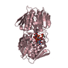
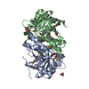
| Title | Crystal Structure of the Lys265Arg PEG-crystallized mutant of monomeric sarcosine oxidase | ||||||
 Components Components | Monomeric sarcosine oxidase | ||||||
 Keywords Keywords | OXIDOREDUCTASE / FLAVOPROTEIN OXIDASE / FAD / Flavoprotein | ||||||
| Function / homology |  Function and homology information Function and homology informationsarcosine oxidase (formaldehyde-forming) / sarcosine oxidase activity / flavin adenine dinucleotide binding / cytoplasm Similarity search - Function | ||||||
| Biological species |  | ||||||
| Method |  X-RAY DIFFRACTION / SYNCHROTRON / MOLECULAR REPLACEMENT / Resolution: 2.1 Å X-RAY DIFFRACTION / SYNCHROTRON / MOLECULAR REPLACEMENT / Resolution: 2.1 Å | ||||||
 Authors Authors | Mathews, F.S. / Chen, Z.-W. / Jorns, M.S. | ||||||
 Citation Citation | Journal: Biochemistry / Year: 2010 Title: Structural characterization of mutations at the oxygen activation site in monomeric sarcosine oxidase. Authors: Jorns, M.S. / Chen, Z.W. / Mathews, F.S. | ||||||
| History |
|
- Structure visualization
| Structure viewer | Molecule: MolmilJmol/JSmol |
|---|
- Downloads & links
-Download
| PDBx/mmCIF format | 3m13.cif.gz | 317.7 KB | Display | PDBx/mmCIF format |
|---|---|---|---|---|
| PDB format | pdb3m13.ent.gz | 258.8 KB | Display | PDB format |
| PDBx/mmJSON format | 3m13.json.gz | Tree view | PDBx/mmJSON format | |
| Others |  Other downloads Other downloads |
-Validation report
| Arichive directory | https://data.pdbj.org/pub/pdb/validation_reports/m1/3m13ftp://data.pdbj.org/pub/pdb/validation_reports/m1/3m13 | HTTPS FTP |
|---|
-Related structure data


-Links
 PDBj
PDBj- Assembly
| Deposited unit | 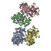
| ||||||||
|---|---|---|---|---|---|---|---|---|---|
| 1 | 
| ||||||||
| 2 | 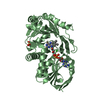
| ||||||||
| 3 | 
| ||||||||
| 4 | 
| ||||||||
| Unit cell |
|
-Components
| #1: Protein | Mass: 43129.371 Da / Num. of mol.: 4 / Mutation: K256R Source method: isolated from a genetically manipulated source Source: (gene. exp.) References: UniProt: P40859, sarcosine oxidase (formaldehyde-forming) #2: Chemical | ChemComp-FAD /   Mass: 785.550 Da / Num. of mol.: 4 / Source method: obtained synthetically / Formula: C27H33N9O15P2 / Comment: FAD*YM Mass: 785.550 Da / Num. of mol.: 4 / Source method: obtained synthetically / Formula: C27H33N9O15P2 / Comment: FAD*YM#3: Chemical | ChemComp-CL /   Mass: 35.453 Da / Num. of mol.: 4 / Source method: obtained synthetically / Formula: Cl Mass: 35.453 Da / Num. of mol.: 4 / Source method: obtained synthetically / Formula: Cl#4: Chemical |   Mass: 94.971 Da / Num. of mol.: 2 / Source method: obtained synthetically / Formula: PO4 Mass: 94.971 Da / Num. of mol.: 2 / Source method: obtained synthetically / Formula: PO4#5: Water | ChemComp-HOH / |  Mass: 18.015 Da / Num. of mol.: 497 / Source method: isolated from a natural source / Formula: H2O Mass: 18.015 Da / Num. of mol.: 497 / Source method: isolated from a natural source / Formula: H2O |
|---|
-Experimental details
-Experiment
| Experiment | Method: X-RAY DIFFRACTION / Number of used crystals: 1 |
|---|
- Sample preparation
| Crystal | Density Matthews: 2.27 Å3/Da / Density % sol: 45.71 % |
|---|---|
| Crystal grow | Temperature: 298 K / Method: vapor diffusion / pH: 8.5 Details: 20% PEG4000, 200 mM sodium acetate, 100 mM Tris HCL, pH 8.5, vapor diffusion, temperature 298K |
-Data collection
| Diffraction | Mean temperature: 100 K |
|---|---|
| Diffraction source | Source: SYNCHROTRON / Site: APS  / Beamline: 14-BM-C / Wavelength: 0.9 Å / Beamline: 14-BM-C / Wavelength: 0.9 Å |
| Detector | Type: ADSC QUANTUM 315 / Detector: CCD |
| Radiation | Protocol: SINGLE WAVELENGTH / Scattering type: x-ray |
| Radiation wavelength | Wavelength: 0.9 Å / Relative weight: 1 |
| Reflection | Resolution: 2→40 Å / Num. obs: 83978 / % possible obs: 95.3 % / Redundancy: 5.9 % / Rmerge(I) obs: 0.072 / Net I/σ(I): 21 |
| Reflection shell | Resolution: 2→2.18 Å / Redundancy: 5.1 % / Rmerge(I) obs: 0.475 / Mean I/σ(I) obs: 3.2 / % possible all: 96 |
- Processing
| Software |
| |||||||||||||||||||||||||||||||||||||||||||||||||||||||||||||||||
|---|---|---|---|---|---|---|---|---|---|---|---|---|---|---|---|---|---|---|---|---|---|---|---|---|---|---|---|---|---|---|---|---|---|---|---|---|---|---|---|---|---|---|---|---|---|---|---|---|---|---|---|---|---|---|---|---|---|---|---|---|---|---|---|---|---|---|
| Refinement | Method to determine structure: MOLECULAR REPLACEMENT / Resolution: 2.1→29.2 Å / Cor.coef. Fo:Fc: 0.959 / Cor.coef. Fo:Fc free: 0.926 / Occupancy max: 1 / Occupancy min: 1 / SU B: 5.609 / SU ML: 0.151 / Cross valid method: THROUGHOUT / σ(F): 0 / ESU R: 0.268 / ESU R Free: 0.215 / Stereochemistry target values: MAXIMUM LIKELIHOOD / Details: HYDROGENS HAVE BEEN ADDED IN THE RIDING POSITIONS
| |||||||||||||||||||||||||||||||||||||||||||||||||||||||||||||||||
| Solvent computation | Ion probe radii: 0.8 Å / Shrinkage radii: 0.8 Å / VDW probe radii: 1.2 Å / Solvent model: MASK | |||||||||||||||||||||||||||||||||||||||||||||||||||||||||||||||||
| Displacement parameters | Biso max: 84.73 Å2 / Biso mean: 37.122 Å2 / Biso min: 10.99 Å2
| |||||||||||||||||||||||||||||||||||||||||||||||||||||||||||||||||
| Refinement step | Cycle: LAST / Resolution: 2.1→29.2 Å
| |||||||||||||||||||||||||||||||||||||||||||||||||||||||||||||||||
| Refine LS restraints |
| |||||||||||||||||||||||||||||||||||||||||||||||||||||||||||||||||
| LS refinement shell | Resolution: 2.101→2.156 Å / Total num. of bins used: 20
|
