[English] 日本語
Yorodumi
- PDB-6m8z: Crystal structure of human DJ-1 without a modification on Cys-106 -

+
Open data


ID or keywords:

Loading...

-
Basic information

Entry
Database: PDB / ID: 6m8z
TitleCrystal structure of human DJ-1 without a modification on Cys-106
ComponentsProtein/nucleic acid deglycase DJ-1
KeywordsUNKNOWN FUNCTION / the exact function of this protein is still unknown
Function / homology
Function and homology information


tyrosine 3-monooxygenase activator activity / cellular response to glyoxal / L-dopa decarboxylase activator activity / peptidyl-cysteine deglycation / peptidyl-arginine deglycation / peptidyl-lysine deglycation / protein deglycation, methylglyoxal removal / glutathione deglycation / detoxification of hydrogen peroxide / methylglyoxal catabolic process to lactate ...tyrosine 3-monooxygenase activator activity / cellular response to glyoxal / L-dopa decarboxylase activator activity / peptidyl-cysteine deglycation / peptidyl-arginine deglycation / peptidyl-lysine deglycation / protein deglycation, methylglyoxal removal / glutathione deglycation / detoxification of hydrogen peroxide / methylglyoxal catabolic process to lactate / guanine deglycation, methylglyoxal removal / cellular detoxification of methylglyoxal / regulation of supramolecular fiber organization / negative regulation of death-inducing signaling complex assembly / negative regulation of TRAIL-activated apoptotic signaling pathway / positive regulation of pyrroline-5-carboxylate reductase activity / positive regulation of tyrosine 3-monooxygenase activity / positive regulation of L-dopa biosynthetic process / positive regulation of L-dopa decarboxylase activity / negative regulation of hydrogen peroxide-induced neuron intrinsic apoptotic signaling pathway / glyoxalase (glycolic acid-forming) activity / negative regulation of protein K48-linked deubiquitination / negative regulation of ubiquitin-specific protease activity / protein deglycation, glyoxal removal / glycolate biosynthetic process / guanine deglycation, glyoxal removal / glyoxal metabolic process / negative regulation of nitrosative stress-induced intrinsic apoptotic signaling pathway / detection of oxidative stress / guanine deglycation / detoxification of mercury ion / protein deglycase / methylglyoxal metabolic process / positive regulation of mitochondrial electron transport, NADH to ubiquinone / mercury ion binding / oxidoreductase activity, acting on peroxide as acceptor / protein deglycase activity / positive regulation of acute inflammatory response to antigenic stimulus / positive regulation of dopamine biosynthetic process / superoxide dismutase copper chaperone activity / positive regulation of NAD(P)H oxidase activity / positive regulation of autophagy of mitochondrion / lactate biosynthetic process / negative regulation of cysteine-type endopeptidase activity involved in apoptotic signaling pathway / cellular detoxification of aldehyde / positive regulation of superoxide dismutase activity / small protein activating enzyme binding / Hydrolases; Acting on ester bonds; Thioester hydrolases / regulation of oxidative stress-induced neuron intrinsic apoptotic signaling pathway / negative regulation of ubiquitin-protein transferase activity / peroxiredoxin activity / detoxification of copper ion / negative regulation of protein acetylation / negative regulation of oxidative stress-induced neuron intrinsic apoptotic signaling pathway / positive regulation of transcription regulatory region DNA binding / positive regulation of oxidative stress-induced intrinsic apoptotic signaling pathway / positive regulation of androgen receptor activity / membrane hyperpolarization / protein deglycosylation / negative regulation of protein sumoylation / oxygen sensor activity / regulation of androgen receptor signaling pathway / negative regulation of protein export from nucleus / negative regulation of intrinsic apoptotic signaling pathway in response to hydrogen peroxide / cupric ion binding / ubiquitin-like protein conjugating enzyme binding / insulin secretion / Hydrolases; Acting on carbon-nitrogen bonds, other than peptide bonds; In linear amides / dopamine uptake involved in synaptic transmission / positive regulation of reactive oxygen species biosynthetic process / nuclear androgen receptor binding / hydrogen peroxide metabolic process / ubiquitin-specific protease binding / cytokine binding / cuprous ion binding / single fertilization / membrane depolarization / negative regulation of proteasomal ubiquitin-dependent protein catabolic process / negative regulation of endoplasmic reticulum stress-induced intrinsic apoptotic signaling pathway / regulation of neuron apoptotic process / negative regulation of oxidative stress-induced intrinsic apoptotic signaling pathway / negative regulation of reactive oxygen species biosynthetic process / negative regulation of protein ubiquitination / activation of protein kinase B activity / mitochondrion organization / adult locomotory behavior / SUMOylation of transcription cofactors / regulation of mitochondrial membrane potential / negative regulation of protein phosphorylation / negative regulation of protein binding / positive regulation of interleukin-8 production / negative regulation of extrinsic apoptotic signaling pathway / positive regulation of protein-containing complex assembly / adherens junction / Late endosomal microautophagy / negative regulation of protein kinase activity / mitochondrial intermembrane space / PML body / cellular response to hydrogen peroxide / autophagy
Similarity search - Function
Protein/nucleic acid deglycase DJ-1 / DJ-1/PfpI / DJ-1/PfpI family / Class I glutamine amidotransferase (GATase) domain / Class I glutamine amidotransferase-like / Rossmann fold / 3-Layer(aba) Sandwich / Alpha Beta
Similarity search - Domain/homology
Parkinson disease protein 7
Similarity search - Component
Biological speciesHomo sapiens (human)
MethodX-RAY DIFFRACTION / SYNCHROTRON / FOURIER SYNTHESIS / Resolution: 1.83 Å
AuthorsShumilin, I.A. / Shabalin, I.G. / Shumilina, S.V. / Werenskjold, C. / Utepbergenov, D. / Minor, W.
Funding support United States, 1items
OrganizationGrant numberCountry
National Institutes of Health/National Institute Of Allergy and Infectious Diseases (NIH/NIAID) United States
CitationJournal: Biochem. Biophys. Res. Commun. / Year: 2018
Title: A transient post-translational modification of active site cysteine alters binding properties of the parkinsonism protein DJ-1.
Authors: Mussakhmetov, A. / Shumilin, I.A. / Nugmanova, R. / Shabalin, I.G. / Baizhumanov, T. / Toibazar, D. / Khassenov, B. / Minor, W. / Utepbergenov, D.
History
DepositionAug 22, 2018Deposition site: RCSB / Processing site: RCSB
Revision 1.0Sep 5, 2018Provider: repository / Type: Initial release
Revision 1.1Sep 19, 2018Group: Data collection / Database references / Category: citation / citation_author
Item: _citation.country / _citation.journal_abbrev ..._citation.country / _citation.journal_abbrev / _citation.journal_id_ASTM / _citation.journal_id_CSD / _citation.journal_id_ISSN / _citation.journal_volume / _citation.page_first / _citation.page_last / _citation.pdbx_database_id_DOI / _citation.pdbx_database_id_PubMed / _citation.title / _citation.year
Revision 1.2Apr 17, 2019Group: Author supporting evidence / Data collection / Category: pdbx_audit_support
Revision 1.3Dec 18, 2019Group: Author supporting evidence / Category: pdbx_audit_support / Item: _pdbx_audit_support.funding_organization
Revision 1.4Apr 13, 2022Group: Database references / Structure summary / Category: audit_author / citation_author / database_2
Item: _audit_author.identifier_ORCID / _citation_author.identifier_ORCID ..._audit_author.identifier_ORCID / _citation_author.identifier_ORCID / _database_2.pdbx_DOI / _database_2.pdbx_database_accession
Revision 1.5Oct 11, 2023Group: Data collection / Refinement description
Category: chem_comp_atom / chem_comp_bond / pdbx_initial_refinement_model

-
Structure visualization

Structure viewerMolecule:
MolmilJmol/JSmol

Downloads & links

-
Assembly

Deposited unit
A: Protein/nucleic acid deglycase DJ-1
hetero molecules


Theoretical massNumber of molelcules
Total (without water)20,3193
Polymers20,0451
Non-polymers2742
Water3,207178
1
A: Protein/nucleic acid deglycase DJ-1
hetero molecules

A: Protein/nucleic acid deglycase DJ-1
hetero molecules


Theoretical massNumber of molelcules
Total (without water)40,6386
Polymers40,0902
Non-polymers5484
Water362
TypeNameSymmetry operationNumber
identity operation1_555x,y,z1
crystal symmetry operation12_555x,x-y,-z+5/61
Buried area3590 Å2
ΔGint-36 kcal/mol
Surface area14730 Å2
MethodPISA
Unit cell
Length a, b, c (Å)66.500, 66.500, 176.728
Angle α, β, γ (deg.)90.000, 90.000, 120.000
Int Tables number179
Space group name H-MP6522
Components on special symmetry positions
IDModelComponents
11A-806-

HOH

21A-869-

HOH

-
Components

#1: Protein Protein/nucleic acid deglycase DJ-1 / Maillard deglycase / Oncogene DJ1 / Parkinson disease protein 7 / Parkinsonism-associated deglycase ...Maillard deglycase / Oncogene DJ1 / Parkinson disease protein 7 / Parkinsonism-associated deglycase / Protein DJ-1 / DJ-1


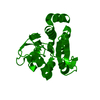
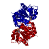
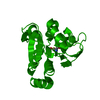
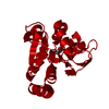
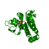
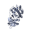
Mass: 20045.180 Da / Num. of mol.: 1
Source method: isolated from a genetically manipulated source
Source: (gene. exp.) Homo sapiens (human) / Gene: PARK7 / Plasmid: pHisParallel / Production host: Escherichia coli (E. coli) / Strain (production host): BL21(DE3) RIPL
References: UniProt: Q99497, Hydrolases; Acting on ester bonds; Thioester hydrolases, Hydrolases; Acting on carbon-nitrogen bonds, other than peptide bonds; In linear amides, protein deglycase
#2: Chemical ChemComp-EPE / 4-(2-HYDROXYETHYL)-1-PIPERAZINE ETHANESULFONIC ACID / HEPES / HEPES


Mass: 238.305 Da / Num. of mol.: 1 / Source method: obtained synthetically / Formula: C8H18N2O4S / Comment: pH buffer*YM
#3: Chemical ChemComp-CL / CHLORIDE ION / Chloride


Mass: 35.453 Da / Num. of mol.: 1 / Source method: obtained synthetically / Formula: Cl
#4: Water ChemComp-HOH / water / Water


Mass: 18.015 Da / Num. of mol.: 178 / Source method: isolated from a natural source / Formula: H2O

-
Experimental details

-
Experiment

ExperimentMethod: X-RAY DIFFRACTION / Number of used crystals: 1

-
Sample preparation

CrystalDensity Matthews: 2.87 Å3/Da / Density % sol: 57.1 %
Crystal growTemperature: 298 K / Method: vapor diffusion, sitting drop / pH: 5.5
Details: 500 nL of protein in 50 mM Tris pH 8.0 and 500 mM NaCl were mixed with 500nL of 0.1M Bis-tris pH 5.5, 0.1M KSCN, and 29% PEG MME 2000

-
Data collection

DiffractionMean temperature: 100 K
Diffraction sourceSource: SYNCHROTRON / Site: APS / Beamline: 21-ID-F / Wavelength: 0.97872 Å
DetectorType: MARMOSAIC 225 mm CCD / Detector: CCD / Date: Jul 6, 2011
RadiationProtocol: SINGLE WAVELENGTH / Monochromatic (M) / Laue (L): M / Scattering type: x-ray
Radiation wavelengthWavelength: 0.97872 Å / Relative weight: 1
ReflectionResolution: 1.83→50 Å / Num. obs: 20934 / % possible obs: 97.8 % / Redundancy: 20.2 % / Rmerge(I) obs: 0.055 / Rpim(I) all: 0.012 / Rrim(I) all: 0.056 / Χ2: 1.087 / Net I/σ(I): 11 / Num. measured all: 423474
Reflection shell

Diffraction-ID: 1

Resolution (Å)Redundancy (%)Rmerge(I) obsNum. unique obsCC1/2Rpim(I) allRrim(I) allΧ2% possible all
1.83-1.8615.81.2488300.5960.3241.2940.80579.3
1.86-1.916.11.0549280.6320.271.0920.79890
1.9-1.9317.70.8210130.8660.1990.8460.82998.3
1.93-1.9720.50.65810280.9350.1470.6740.88898.9
1.97-2.0121.50.53910490.9620.1170.5520.81199.2
2.01-2.0621.40.43810250.9760.0950.4480.85199.2
2.06-2.1121.50.32210350.9840.070.330.90699.2
2.11-2.1721.50.27110450.990.0590.2780.9499.3
2.17-2.2321.30.20710390.9940.0450.2120.96199.5
2.23-2.3121.40.17810540.9950.0390.1821.02699.4
2.31-2.3921.30.14710430.9970.0320.1511.07299.5
2.39-2.4821.40.12410520.9970.0270.1271.11799.5
2.48-2.621.20.10310540.9970.0230.1061.299.7
2.6-2.7321.20.08510630.9990.0190.0871.26699.6
2.73-2.9210.06610810.9990.0150.0681.31599.6
2.9-3.1320.90.05610610.9990.0120.0571.43599.6
3.13-3.4420.40.04610850.9990.010.0471.48799.9
3.44-3.94200.04111110.9990.0090.0421.59199.7
3.94-4.9719.60.03411240.9990.0080.0351.36599.5
4.97-5017.80.025121410.0060.0260.80196.4

-
Processing

Software
NameVersionClassification
HKL-3000data scaling
REFMAC5.8.0230refinement
PDB_EXTRACT3.24data extraction
HKL-3000data reduction
HKL-3000phasing
RefinementMethod to determine structure: FOURIER SYNTHESIS
Starting model: 6E5Z
Resolution: 1.83→32.7 Å / Cor.coef. Fo:Fc: 0.963 / Cor.coef. Fo:Fc free: 0.951 / SU B: 5.264 / SU ML: 0.08 / SU R Cruickshank DPI: 0.112 / Cross valid method: THROUGHOUT / σ(F): 0 / ESU R: 0.112 / ESU R Free: 0.11
Details: U VALUES : WITH TLS ADDED HYDROGENS HAVE BEEN ADDED IN THE RIDING POSITIONS
RfactorNum. reflection% reflectionSelection details
Rfree0.2043 1051 5.2 %RANDOM
Rwork0.1699 ---
obs0.1718 19231 95.27 %-
Solvent computationIon probe radii: 0.8 Å / Shrinkage radii: 0.8 Å / VDW probe radii: 1.2 Å
Displacement parametersBiso max: 94.52 Å2 / Biso mean: 29.366 Å2 / Biso min: 7.34 Å2
Baniso -1Baniso -2Baniso -3
1--0.4 Å2-0.2 Å2-0 Å2
2---0.4 Å20 Å2
3---1.29 Å2
Refinement stepCycle: final / Resolution: 1.83→32.7 Å
ProteinNucleic acidLigandSolventTotal
Num. atoms1375 0 8 178 1561
Biso mean--42.35 40.61 -
Num. residues----187
Refine LS restraints
Refine-IDTypeDev idealDev ideal targetNumber
X-RAY DIFFRACTIONr_bond_refined_d0.0070.0141408
X-RAY DIFFRACTIONr_bond_other_d0.0010.0171377
X-RAY DIFFRACTIONr_angle_refined_deg1.1641.6561904
X-RAY DIFFRACTIONr_angle_other_deg0.8771.6443226
X-RAY DIFFRACTIONr_dihedral_angle_1_deg6.4945190
X-RAY DIFFRACTIONr_dihedral_angle_2_deg33.67623.39356
X-RAY DIFFRACTIONr_dihedral_angle_3_deg13.56715256
X-RAY DIFFRACTIONr_dihedral_angle_4_deg16.927157
X-RAY DIFFRACTIONr_chiral_restr0.0590.2190
X-RAY DIFFRACTIONr_gen_planes_refined0.0050.021565
X-RAY DIFFRACTIONr_gen_planes_other0.0010.02219
LS refinement shellResolution: 1.83→1.877 Å / Rfactor Rfree error: 0 / Total num. of bins used: 20
RfactorNum. reflection% reflection
Rfree0.233 50 -
Rwork0.243 920 -
all-970 -
obs--63.69 %
Refinement TLS params.Method: refined / Origin x: 28.435 Å / Origin y: 2.224 Å / Origin z: 67.191 Å
111213212223313233
T0.1867 Å20.1005 Å20.0574 Å2-0.0906 Å20.0317 Å2--0.0656 Å2
L1.3109 °20.2043 °2-0.2471 °2-1.5009 °2-0.3253 °2--3.9134 °2
S-0.084 Å °0.1472 Å °-0.1329 Å °-0.139 Å °-0.1546 Å °-0.2781 Å °0.6317 Å °0.352 Å °0.2386 Å °

+
About Yorodumi

-
News

-
Feb 9, 2022. New format data for meta-information of EMDB entries

New format data for meta-information of EMDB entries

  • Version 3 of the EMDB header file is now the official format.
  • The previous official version 1.9 will be removed from the archive.

Related info.:EMDB header

External links:wwPDB to switch to version 3 of the EMDB data model

-
Aug 12, 2020. Covid-19 info

Covid-19 info

URL: https://pdbj.org/emnavi/covid19.php

New page: Covid-19 featured information page in EM Navigator.

Related info.:Covid-19 info / Mar 5, 2020. Novel coronavirus structure data

+
Mar 5, 2020. Novel coronavirus structure data

Novel coronavirus structure data

Related info.:Yorodumi Speices / Aug 12, 2020. Covid-19 info

External links:COVID-19 featured content - PDBj / Molecule of the Month (242):Coronavirus Proteases

+
Jan 31, 2019. EMDB accession codes are about to change! (news from PDBe EMDB page)

EMDB accession codes are about to change! (news from PDBe EMDB page)

  • The allocation of 4 digits for EMDB accession codes will soon come to an end. Whilst these codes will remain in use, new EMDB accession codes will include an additional digit and will expand incrementally as the available range of codes is exhausted. The current 4-digit format prefixed with “EMD-” (i.e. EMD-XXXX) will advance to a 5-digit format (i.e. EMD-XXXXX), and so on. It is currently estimated that the 4-digit codes will be depleted around Spring 2019, at which point the 5-digit format will come into force.
  • The EM Navigator/Yorodumi systems omit the EMD- prefix.

Related info.:Q: What is EMD? / ID/Accession-code notation in Yorodumi/EM Navigator

External links:EMDB Accession Codes are Changing Soon! / Contact to PDBj

+
Jul 12, 2017. Major update of PDB

Major update of PDB

  • wwPDB released updated PDB data conforming to the new PDBx/mmCIF dictionary.
  • This is a major update changing the version number from 4 to 5, and with Remediation, in which all the entries are updated.
  • In this update, many items about electron microscopy experimental information are reorganized (e.g. em_software).
  • Now, EM Navigator and Yorodumi are based on the updated data.

External links:wwPDB Remediation / Enriched Model Files Conforming to OneDep Data Standards Now Available in the PDB FTP Archive

-
Yorodumi

Thousand views of thousand structures

  • Yorodumi is a browser for structure data from EMDB, PDB, SASBDB, etc.
  • This page is also the successor to EM Navigator detail page, and also detail information page/front-end page for Omokage search.
  • The word "yorodu" (or yorozu) is an old Japanese word meaning "ten thousand". "mi" (miru) is to see.

Related info.:EMDB / PDB / SASBDB / Comparison of 3 databanks / Yorodumi Search / Aug 31, 2016. New EM Navigator & Yorodumi / Yorodumi Papers / Jmol/JSmol / Function and homology information / Changes in new EM Navigator and Yorodumi

Read more