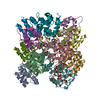
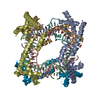
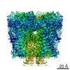
Journal: Proc Natl Acad Sci U S A / Year: 2015 Title: Structure and assembly of the mouse ASC inflammasome by combined NMR spectroscopy and cryo-electron microscopy. Authors: Lorenzo Sborgi / Francesco Ravotti / Venkata P Dandey / Mathias S Dick / Adam Mazur / Sina Reckel / Mohamed Chami / Sebastian Scherer / Matthias Huber / Anja Böckmann / Edward H Egelman / ...Authors: Lorenzo Sborgi / Francesco Ravotti / Venkata P Dandey / Mathias S Dick / Adam Mazur / Sina Reckel / Mohamed Chami / Sebastian Scherer / Matthias Huber / Anja Böckmann / Edward H Egelman / Henning Stahlberg / Petr Broz / Beat H Meier / Sebastian Hiller / Abstract: Inflammasomes are multiprotein complexes that control the innate immune response by activating caspase-1, thus promoting the secretion of cytokines in response to invading pathogens and endogenous ...Inflammasomes are multiprotein complexes that control the innate immune response by activating caspase-1, thus promoting the secretion of cytokines in response to invading pathogens and endogenous triggers. Assembly of inflammasomes is induced by activation of a receptor protein. Many inflammasome receptors require the adapter protein ASC [apoptosis-associated speck-like protein containing a caspase-recruitment domain (CARD)], which consists of two domains, the N-terminal pyrin domain (PYD) and the C-terminal CARD. Upon activation, ASC forms large oligomeric filaments, which facilitate procaspase-1 recruitment. Here, we characterize the structure and filament formation of mouse ASC in vitro at atomic resolution. Information from cryo-electron microscopy and solid-state NMR spectroscopy is combined in a single structure calculation to obtain the atomic-resolution structure of the ASC filament. Perturbations of NMR resonances upon filament formation monitor the specific binding interfaces of ASC-PYD association. Importantly, NMR experiments show the rigidity of the PYD forming the core of the filament as well as the high mobility of the CARD relative to this core. The findings are validated by structure-based mutagenesis experiments in cultured macrophages. The 3D structure of the mouse ASC-PYD filament is highly similar to the recently determined human ASC-PYD filament, suggesting evolutionary conservation of ASC-dependent inflammasome mechanisms.
A: Apoptosis-associated speck-like protein B: Apoptosis-associated speck-like protein C: Apoptosis-associated speck-like protein D: Apoptosis-associated speck-like protein E: Apoptosis-associated speck-like protein F: Apoptosis-associated speck-like protein G: Apoptosis-associated speck-like protein H: Apoptosis-associated speck-like protein I: Apoptosis-associated speck-like protein J: Apoptosis-associated speck-like protein K: Apoptosis-associated speck-like protein L: Apoptosis-associated speck-like protein M: Apoptosis-associated speck-like protein N: Apoptosis-associated speck-like protein O: Apoptosis-associated speck-like protein
Helical symmetry: (Circular symmetry: 3 / Dyad axis: no / N subunits divisor: 1 / Num. of operations: 8 / Rise per n subunits: 14.2 Å / Rotation per n subunits: 53 °)
Details
The helical parameters generate the filament from any single chain.
-
Components
#1: Protein
Apoptosis-associatedspeck-likeprotein / mASC
Mass: 10118.684 Da / Num. of mol.: 15 / Fragment: pyrin domain (UNP residues 2-90) Source method: isolated from a genetically manipulated source Source: (gene. exp.) Mus musculus (house mouse) / Gene: Pycard, Asc / Plasmid: pET28a / Production host: Escherichia coli BL21(DE3) (bacteria) / References: UniProt: Q9EPB4
-
Experimental details
-
Experiment
Experiment
Method
SOLID-STATE NMR
ELECTRON MICROSCOPY
EM experiment
Aggregation state: FILAMENT / 3D reconstruction method: helical reconstruction
NMR experiment
Conditions-ID
Experiment-ID
Solution-ID
Type
1
1
1
NCACX
1
2
1
NCOCX
1
3
1
NCACB
1
4
1
CANCO
1
5
1
CCC
1
6
1
NCA
1
7
1
NCO
1
8
1
20msDARR
1
9
1
N(CO)CACB
1
10
1
CAN(CO)CA
1
11
1
N(CA)CBCX
1
12
1
100msPDSD
-
Sample preparation
Component
ID
Name
Type
Parent-ID
1
Mouse ASC-PYD filament
COMPLEX
0
2
ASCfilament
1
Buffer solution
Name: 25 mM Tris, 300 mM sodium chloride / pH: 8 / Details: 25 mM Tris, 300 mM sodium chloride
Specimen
Embedding applied: NO / Shadowing applied: NO / Staining applied: NO / Vitrification applied: YES
Vitrification
Instrument: FEI VITROBOT MARK IV / Cryogen name: ETHANE Details: Grids were blotted for one second before plunging into liquid ethane (FEI VITROBOT MARK IV).
In the structure databanks used in Yorodumi, some data are registered as the other names, "COVID-19 virus" and "2019-nCoV". Here are the details of the virus and the list of structure data.
Jan 31, 2019. EMDB accession codes are about to change! (news from PDBe EMDB page)
EMDB accession codes are about to change! (news from PDBe EMDB page)
The allocation of 4 digits for EMDB accession codes will soon come to an end. Whilst these codes will remain in use, new EMDB accession codes will include an additional digit and will expand incrementally as the available range of codes is exhausted. The current 4-digit format prefixed with “EMD-” (i.e. EMD-XXXX) will advance to a 5-digit format (i.e. EMD-XXXXX), and so on. It is currently estimated that the 4-digit codes will be depleted around Spring 2019, at which point the 5-digit format will come into force.
The EM Navigator/Yorodumi systems omit the EMD- prefix.
Related info.:Q: What is EMD? / ID/Accession-code notation in Yorodumi/EM Navigator
Yorodumi is a browser for structure data from EMDB, PDB, SASBDB, etc.
This page is also the successor to EM Navigator detail page, and also detail information page/front-end page for Omokage search.
The word "yorodu" (or yorozu) is an old Japanese word meaning "ten thousand". "mi" (miru) is to see.
Related info.:EMDB / PDB / SASBDB / Comparison of 3 databanks / Yorodumi Search / Aug 31, 2016. New EM Navigator & Yorodumi / Yorodumi Papers / Jmol/JSmol / Function and homology information / Changes in new EM Navigator and Yorodumi
 Movie
Movie Controller

 Yorodumi
 Open data
 Basic information
 Components
 Keywords
 Function and homology information
 Authors
 Citation


 Structure visualization
 UCSF Chimera
 Downloads & links
 Other downloads


 PDBj
PDBj


 Assembly

 Sample preparation
 Processing