 Movie
Movie Controller

[English] 日本語
 Yorodumi
- PDB-1v1j: Crystal structure of type II Dehydroquintae Dehydratase from Stre... -
+ Open data

- Basic information
| Entry | Database: PDB / ID: 1v1j | ||||||
|---|---|---|---|---|---|---|---|
| Title | Crystal structure of type II Dehydroquintae Dehydratase from Streptomyces coelicolor in complex with 3-fluoro | ||||||
 Components Components | 3-DEHYDROQUINATE DEHYDRATASE | ||||||
 Keywords Keywords | LYASE / DHQ / SHIKIMATE / DEHYDROQUINASE / AROMATIC AMINO ACID BIOSYNTHESIS | ||||||
| Function / homology |  Function and homology information Function and homology informationquinate catabolic process / 3-dehydroquinate dehydratase / 3-dehydroquinate dehydratase activity / chorismate biosynthetic process / aromatic amino acid biosynthetic process / amino acid biosynthetic process Similarity search - Function | ||||||
| Biological species |  STREPTOMYCES COELICOLOR (bacteria) STREPTOMYCES COELICOLOR (bacteria) | ||||||
| Method |  X-RAY DIFFRACTION / MOLECULAR REPLACEMENT / Resolution: 2.2 Å X-RAY DIFFRACTION / MOLECULAR REPLACEMENT / Resolution: 2.2 Å | ||||||
 Authors Authors | Roszak, A.W. / Coggins, J.R. / Lapthorn, A.J. | ||||||
 Citation Citation | Journal: Org.Biomol.Chem. / Year: 2004 Title: (1R,4S,5R)-3-Fluoro-1,4,5-Trihydroxy-2-Cyclohexene-1-Carboxylic Acid: The Fluoro Analogue of the Enolate Intermediate in the Reaction Catalyzed by Type II Dehydroquinases Authors: Frederickson, M. / Roszak, A.W. / Coggins, J.R. / Lapthorn, A.J. / Abell, C. | ||||||
| History |
|
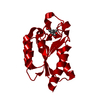
- Structure visualization
| Structure viewer | Molecule: MolmilJmol/JSmol |
|---|
- Downloads & links
-Download
| PDBx/mmCIF format | 1v1j.cif.gz | 354.4 KB | Display | PDBx/mmCIF format |
|---|---|---|---|---|
| PDB format | pdb1v1j.ent.gz | 293.2 KB | Display | PDB format |
| PDBx/mmJSON format | 1v1j.json.gz | Tree view | PDBx/mmJSON format | |
| Others |  Other downloads Other downloads |
-Validation report
| Arichive directory | https://data.pdbj.org/pub/pdb/validation_reports/v1/1v1jftp://data.pdbj.org/pub/pdb/validation_reports/v1/1v1j | HTTPS FTP |
|---|
-Related structure data
| Related structure data |  1guoS S: Starting model for refinement |
|---|---|
| Similar structure data |


-Links
 PDBj
PDBj
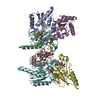
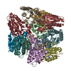
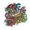
- Assembly
| Deposited unit | 
| ||||||||||||||||||||||||||||||||||||||||||||||||
|---|---|---|---|---|---|---|---|---|---|---|---|---|---|---|---|---|---|---|---|---|---|---|---|---|---|---|---|---|---|---|---|---|---|---|---|---|---|---|---|---|---|---|---|---|---|---|---|---|---|
| 1 |
| ||||||||||||||||||||||||||||||||||||||||||||||||
| Unit cell |
| ||||||||||||||||||||||||||||||||||||||||||||||||
| Noncrystallographic symmetry (NCS) | NCS oper:
|
-Components
| #1: Protein | Mass: 16700.801 Da / Num. of mol.: 12 Source method: isolated from a genetically manipulated source Source: (gene. exp.) STREPTOMYCES COELICOLOR (bacteria) / Plasmid: PTB361 / Production host: #2: Chemical | ChemComp-FA3 / 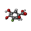  Mass: 192.142 Da / Num. of mol.: 12 / Source method: obtained synthetically / Formula: C7H9FO5 Mass: 192.142 Da / Num. of mol.: 12 / Source method: obtained synthetically / Formula: C7H9FO5#3: Chemical | ChemComp-TRS /   Mass: 122.143 Da / Num. of mol.: 4 / Source method: obtained synthetically / Formula: C4H12NO3 / Comment: pH buffer*YM Mass: 122.143 Da / Num. of mol.: 4 / Source method: obtained synthetically / Formula: C4H12NO3 / Comment: pH buffer*YM#4: Water | ChemComp-HOH / |  Mass: 18.015 Da / Num. of mol.: 700 / Source method: isolated from a natural source / Formula: H2O Mass: 18.015 Da / Num. of mol.: 700 / Source method: isolated from a natural source / Formula: H2O |
|---|
-Experimental details
-Experiment
| Experiment | Method: X-RAY DIFFRACTION / Number of used crystals: 1 |
|---|
- Sample preparation
| Crystal | Density Matthews: 3 Å3/Da / Density % sol: 58.5 % |
|---|---|
| Crystal grow | pH: 7.5 / Details: PEG 8000, NA/K PHOSPHATE, TRIS BUFFER, pH 7.50 |
-Data collection
| Diffraction | Mean temperature: 100 K |
|---|---|
| Diffraction source | Source: ROTATING ANODE / Type: ENRAF-NONIUS FR591 / Wavelength: 1.5418 |
| Detector | Type: MACSCIENCE / Detector: IMAGE PLATE / Date: Oct 15, 2000 / Details: MIRRORS |
| Radiation | Protocol: SINGLE WAVELENGTH / Monochromatic (M) / Laue (L): M / Scattering type: x-ray |
| Radiation wavelength | Wavelength: 1.5418 Å / Relative weight: 1 |
| Reflection | Resolution: 2.2→50 Å / Num. obs: 113346 / % possible obs: 96.6 % / Redundancy: 3.2 % / Biso Wilson estimate: 25.11 Å2 / Rmerge(I) obs: 0.16 / Net I/σ(I): 6.97 |
| Reflection shell | Resolution: 2.2→2.24 Å / Redundancy: 2.5 % / Rmerge(I) obs: 0.77 / Mean I/σ(I) obs: 1.85 / % possible all: 99.5 |
- Processing
| Software |
| ||||||||||||||||||||
|---|---|---|---|---|---|---|---|---|---|---|---|---|---|---|---|---|---|---|---|---|---|
| Refinement | Method to determine structure: MOLECULAR REPLACEMENT Starting model: PDB ENTRY 1GUO Resolution: 2.2→20 Å / SU B: 9.91074 / SU ML: 0.24061 / Cross valid method: THROUGHOUT / σ(F): 0 / ESU R: 0.34862 / ESU R Free: 0.22984
| ||||||||||||||||||||
| Displacement parameters | Biso mean: 25.126 Å2
| ||||||||||||||||||||
| Refinement step | Cycle: LAST / Resolution: 2.2→20 Å
|
