 Movie
Movie Controller


+ Open data

- Basic information
| Entry | Database: PDB / ID: 1guo | ||||||
|---|---|---|---|---|---|---|---|
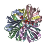
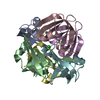
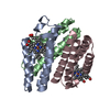
| Title | MopII from Clostridium pasteurianum complexed with molybdate | ||||||
 Components Components | MOLYBDATE BINDING PROTEIN II | ||||||
 Keywords Keywords | TRANSPORT PROTEIN / MOLYBDATE BINDING / MOP / MOLYBDENUM | ||||||
| Function / homology |  Function and homology information Function and homology information | ||||||
| Biological species |  CLOSTRIDIUM PASTEURIANUM (bacteria) CLOSTRIDIUM PASTEURIANUM (bacteria) | ||||||
| Method |  X-RAY DIFFRACTION / MOLECULAR REPLACEMENT / Resolution: 2.5 Å X-RAY DIFFRACTION / MOLECULAR REPLACEMENT / Resolution: 2.5 Å | ||||||
 Authors Authors | Schuettelkopf, A.W. / Harrison, J.A. / Hunter, W.N. | ||||||
 Citation Citation | Journal: J.Biol.Chem. / Year: 2002 Title: Passive Acquisition of Ligand by the Mopii Molbindin from Clostridium Pasteurianum: Structures of Apo and Oxyanion-Bound Forms Authors: Schuettelkopf, A.W. / Harrison, J.A. / Boxer, D.H. / Hunter, W.N. | ||||||
| History |
|
- Structure visualization
| Structure viewer | Molecule: MolmilJmol/JSmol |
|---|
- Downloads & links
-Download
| PDBx/mmCIF format | 1guo.cif.gz | 82.6 KB | Display | PDBx/mmCIF format |
|---|---|---|---|---|
| PDB format | pdb1guo.ent.gz | 64.4 KB | Display | PDB format |
| PDBx/mmJSON format | 1guo.json.gz | Tree view | PDBx/mmJSON format | |
| Others |  Other downloads Other downloads |
-Validation report
| Arichive directory | https://data.pdbj.org/pub/pdb/validation_reports/gu/1guoftp://data.pdbj.org/pub/pdb/validation_reports/gu/1guo | HTTPS FTP |
|---|
-Related structure data
| Related structure data |  1gugSC 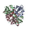 1gunC  1gusC 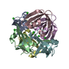 1gutC S: Starting model for refinement C: citing same article ( |
|---|---|
| Similar structure data |


-Links
 PDBj
PDBj- Assembly
| Deposited unit | 
| ||||||||||||||||||||||||
|---|---|---|---|---|---|---|---|---|---|---|---|---|---|---|---|---|---|---|---|---|---|---|---|---|---|
| 1 | 
| ||||||||||||||||||||||||
| 2 | 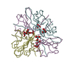
| ||||||||||||||||||||||||
| Unit cell |
| ||||||||||||||||||||||||
| Components on special symmetry positions |
| ||||||||||||||||||||||||
| Noncrystallographic symmetry (NCS) | NCS oper:
|
-Components
| #1: Protein | Mass: 7017.250 Da / Num. of mol.: 6 Source method: isolated from a genetically manipulated source Source: (gene. exp.) CLOSTRIDIUM PASTEURIANUM (bacteria) / Plasmid: PET15B / Production host: #2: Chemical | ChemComp-MOO /   Mass: 159.938 Da / Num. of mol.: 8 / Source method: obtained synthetically / Formula: MoO4 Mass: 159.938 Da / Num. of mol.: 8 / Source method: obtained synthetically / Formula: MoO4#3: Water | ChemComp-HOH / |  Mass: 18.015 Da / Num. of mol.: 13 / Source method: isolated from a natural source / Formula: H2O Mass: 18.015 Da / Num. of mol.: 13 / Source method: isolated from a natural source / Formula: H2O |
|---|
-Experimental details
-Experiment
| Experiment | Method: X-RAY DIFFRACTION / Number of used crystals: 1 |
|---|
- Sample preparation
| Crystal | Density Matthews: 2.4 Å3/Da / Density % sol: 48 % | |||||||||||||||||||||||||||||||||||||||||||||||||
|---|---|---|---|---|---|---|---|---|---|---|---|---|---|---|---|---|---|---|---|---|---|---|---|---|---|---|---|---|---|---|---|---|---|---|---|---|---|---|---|---|---|---|---|---|---|---|---|---|---|---|
| Crystal grow | pH: 7.6 Details: 95 MM HEPES PH 7.5, 27% POLYETHYLENE GLYCOL 400,5% GLYCEROL, 190 MM CACL2 WITH 1.6 MM NA2MOO4 IN THE DROP | |||||||||||||||||||||||||||||||||||||||||||||||||
| Crystal grow | *PLUS Temperature: 293 K / pH: 7.5 / Method: vapor diffusion, hanging dropDetails: Harrison, J.A., (2001) Acta Crystallogr., D57, 1715. | |||||||||||||||||||||||||||||||||||||||||||||||||
| Components of the solutions | *PLUS
|
-Data collection
| Diffraction | Mean temperature: 100 K |
|---|---|
| Diffraction source | Source: ROTATING ANODE / Type: RIGAKU RU200 / Wavelength: 1.54 |
| Detector | Type: RIGAKU IMAGE PLATE / Detector: IMAGE PLATE / Date: Jul 15, 2001 / Details: MIRRORS |
| Radiation | Protocol: SINGLE WAVELENGTH / Monochromatic (M) / Laue (L): M / Scattering type: x-ray |
| Radiation wavelength | Wavelength: 1.54 Å / Relative weight: 1 |
| Reflection | Resolution: 2.4→30 Å / Num. obs: 10097 / % possible obs: 98.3 % / Redundancy: 4 % / Rmerge(I) obs: 0.05 / Net I/σ(I): 27.7 |
| Reflection shell | Resolution: 2.4→2.49 Å / Redundancy: 2.9 % / Rmerge(I) obs: 0.259 / Mean I/σ(I) obs: 4.4 / % possible all: 89.2 |
| Reflection | *PLUS Num. measured all: 173881 / Rmerge(I) obs: 0.05 |
| Reflection shell | *PLUS % possible obs: 89.2 % |
- Processing
| Software |
| ||||||||||||||||||||||||||||||||||||||||||||||||||||||||||||||||||||||||||||||||||||||||||||||||||||||||||||||||||||||||||||||||||||||||||||||||||||||||||||||||||||||||||||||||||||||
|---|---|---|---|---|---|---|---|---|---|---|---|---|---|---|---|---|---|---|---|---|---|---|---|---|---|---|---|---|---|---|---|---|---|---|---|---|---|---|---|---|---|---|---|---|---|---|---|---|---|---|---|---|---|---|---|---|---|---|---|---|---|---|---|---|---|---|---|---|---|---|---|---|---|---|---|---|---|---|---|---|---|---|---|---|---|---|---|---|---|---|---|---|---|---|---|---|---|---|---|---|---|---|---|---|---|---|---|---|---|---|---|---|---|---|---|---|---|---|---|---|---|---|---|---|---|---|---|---|---|---|---|---|---|---|---|---|---|---|---|---|---|---|---|---|---|---|---|---|---|---|---|---|---|---|---|---|---|---|---|---|---|---|---|---|---|---|---|---|---|---|---|---|---|---|---|---|---|---|---|---|---|---|---|
| Refinement | Method to determine structure: MOLECULAR REPLACEMENT Starting model: PDB ENTRY 1GUG Resolution: 2.5→29.88 Å / Cor.coef. Fo:Fc: 0.939 / Cor.coef. Fo:Fc free: 0.92 / SU B: 13.65 / SU ML: 0.31 / Cross valid method: THROUGHOUT / ESU R Free: 0.356 / Stereochemistry target values: MAXIMUM LIKELIHOOD Details: THE DATA SET WAS ORIGINALLY PROCESSED/ SCALED IN AN ORTHORHOMBIC SPACE GROUP, BUT COULD NOT BE REFINED WITH THE ADDITIONAL CRYSTALLOGRAPHIC SYMMETRY.
| ||||||||||||||||||||||||||||||||||||||||||||||||||||||||||||||||||||||||||||||||||||||||||||||||||||||||||||||||||||||||||||||||||||||||||||||||||||||||||||||||||||||||||||||||||||||
| Solvent computation | Ion probe radii: 0.8 Å / Shrinkage radii: 0.8 Å / VDW probe radii: 1.4 Å / Solvent model: BABINET MODEL WITH MASK | ||||||||||||||||||||||||||||||||||||||||||||||||||||||||||||||||||||||||||||||||||||||||||||||||||||||||||||||||||||||||||||||||||||||||||||||||||||||||||||||||||||||||||||||||||||||
| Refinement step | Cycle: LAST / Resolution: 2.5→29.88 Å
| ||||||||||||||||||||||||||||||||||||||||||||||||||||||||||||||||||||||||||||||||||||||||||||||||||||||||||||||||||||||||||||||||||||||||||||||||||||||||||||||||||||||||||||||||||||||
| Refine LS restraints |
|