 Movie
Movie Controller

[English] 日本語
 Yorodumi
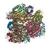
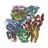
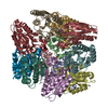
- PDB-1d0i: CRYSTAL STRUCTURE OF TYPE II DEHYDROQUINASE FROM STREPTOMYCES COE... -
+ Open data

- Basic information
| Entry | Database: PDB / ID: 1d0i | ||||||
|---|---|---|---|---|---|---|---|
| Title | CRYSTAL STRUCTURE OF TYPE II DEHYDROQUINASE FROM STREPTOMYCES COELICOLOR COMPLEXED WITH PHOSPHATE IONS | ||||||
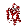
 Components Components | TYPE II 3-DEHYDROQUINATE HYDRATASE | ||||||
 Keywords Keywords | LYASE / TYPE II DEHYDROQUINASE / SHIKIMATE PATHWAY / DODECAMERIC QUATERNARY STRUCTURE / TETRAHEDRAL SYMMETRY | ||||||
| Function / homology |  Function and homology information Function and homology informationquinate catabolic process / 3-dehydroquinate dehydratase / 3-dehydroquinate dehydratase activity / chorismate biosynthetic process / aromatic amino acid biosynthetic process / amino acid biosynthetic process Similarity search - Function | ||||||
| Biological species |  Streptomyces coelicolor (bacteria) Streptomyces coelicolor (bacteria) | ||||||
| Method |  X-RAY DIFFRACTION / SYNCHROTRON / Resolution: 1.8 Å X-RAY DIFFRACTION / SYNCHROTRON / Resolution: 1.8 Å | ||||||
 Authors Authors | Roszak, A.W. / Krell, T. / Hunter, I.S. / Coggins, J.R. / Lapthorn, A.J. | ||||||
 Citation Citation | Journal: Structure / Year: 2002 Title: The structure and mechanism of the type II dehydroquinase from Streptomyces coelicolor. Authors: Roszak, A.W. / Robinson, D.A. / Krell, T. / Hunter, I.S. / Fredrickson, M. / Abell, C. / Coggins, J.R. / Lapthorn, A.J. | ||||||
| History |
|
- Structure visualization
| Structure viewer | Molecule: MolmilJmol/JSmol |
|---|
- Downloads & links
-Download
| PDBx/mmCIF format | 1d0i.cif.gz | 758.4 KB | Display | PDBx/mmCIF format |
|---|---|---|---|---|
| PDB format | pdb1d0i.ent.gz | 629.7 KB | Display | PDB format |
| PDBx/mmJSON format | 1d0i.json.gz | Tree view | PDBx/mmJSON format | |
| Others |  Other downloads Other downloads |
-Validation report
| Arichive directory | https://data.pdbj.org/pub/pdb/validation_reports/d0/1d0iftp://data.pdbj.org/pub/pdb/validation_reports/d0/1d0i | HTTPS FTP |
|---|
-Related structure data


-Links
 PDBj
PDBj
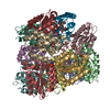
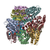
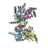
- Assembly
| Deposited unit | 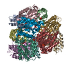
| ||||||||
|---|---|---|---|---|---|---|---|---|---|
| 1 |
| ||||||||
| Unit cell |
|
-Components
| #1: Protein | Mass: 16569.605 Da / Num. of mol.: 12 Source method: isolated from a genetically manipulated source Source: (gene. exp.) Streptomyces coelicolor (bacteria) / Description: STREPTOMYCES COELICOLOR / Plasmid: PDHQ / Production host: #2: Chemical | ChemComp-PO4 /   Mass: 94.971 Da / Num. of mol.: 16 / Source method: obtained synthetically / Formula: PO4 Mass: 94.971 Da / Num. of mol.: 16 / Source method: obtained synthetically / Formula: PO4#3: Chemical | ChemComp-TRS /   Mass: 122.143 Da / Num. of mol.: 4 / Source method: obtained synthetically / Formula: C4H12NO3 / Comment: pH buffer*YM Mass: 122.143 Da / Num. of mol.: 4 / Source method: obtained synthetically / Formula: C4H12NO3 / Comment: pH buffer*YM#4: Water | ChemComp-HOH / |  Mass: 18.015 Da / Num. of mol.: 2023 / Source method: isolated from a natural source / Formula: H2O Mass: 18.015 Da / Num. of mol.: 2023 / Source method: isolated from a natural source / Formula: H2O |
|---|
-Experimental details
-Experiment
| Experiment | Method: X-RAY DIFFRACTION / Number of used crystals: 1 |
|---|
- Sample preparation
| Crystal | Density Matthews: 2.74 Å3/Da / Density % sol: 55.15 % | ||||||||||||||||||||
|---|---|---|---|---|---|---|---|---|---|---|---|---|---|---|---|---|---|---|---|---|---|
| Crystal grow | Temperature: 295 K / Method: vapor diffusion, sitting drop / pH: 8.5 Details: PEG 8000, SODIUM/POTASSIUM PHOSPHATE, TRIS BUFFER, pH 8.5, VAPOR DIFFUSION, SITTING DROP, temperature 295K | ||||||||||||||||||||
| Crystal | *PLUS Density % sol: 57.1 % | ||||||||||||||||||||
| Crystal grow | *PLUS Method: unknown | ||||||||||||||||||||
| Components of the solutions | *PLUS
|
-Data collection
| Diffraction | Mean temperature: 100 K |
|---|---|
| Diffraction source | Source: SYNCHROTRON / Site: SRS  / Beamline: PX9.6 / Wavelength: 0.89 / Beamline: PX9.6 / Wavelength: 0.89 |
| Detector | Type: ADSC QUANTUM 4 / Detector: CCD / Date: Mar 4, 1998 |
| Radiation | Protocol: SINGLE WAVELENGTH / Monochromatic (M) / Laue (L): M / Scattering type: x-ray |
| Radiation wavelength | Wavelength: 0.89 Å / Relative weight: 1 |
| Reflection | Resolution: 1.8→45.03 Å / Num. all: 199360 / Num. obs: 199360 / % possible obs: 98.7 % / Observed criterion σ(F): 0 / Observed criterion σ(I): 0 / Redundancy: 4 % / Biso Wilson estimate: 23.5 Å2 / Rmerge(I) obs: 0.053 / Net I/σ(I): 13.6 |
| Reflection shell | Resolution: 1.8→1.82 Å / Redundancy: 2.9 % / Rmerge(I) obs: 0.593 / % possible all: 93.5 |
| Reflection | *PLUS Num. measured all: 798417 |
| Reflection shell | *PLUS % possible obs: 93.5 % |
- Processing
| Software |
| |||||||||||||||||||||||||||||||||||||||||||||||||||||||||||||||
|---|---|---|---|---|---|---|---|---|---|---|---|---|---|---|---|---|---|---|---|---|---|---|---|---|---|---|---|---|---|---|---|---|---|---|---|---|---|---|---|---|---|---|---|---|---|---|---|---|---|---|---|---|---|---|---|---|---|---|---|---|---|---|---|---|
| Refinement | Resolution: 1.8→45.03 Å / σ(F): 0 / Stereochemistry target values: ENGH & HUBER Details: USED RESTRAINED REFINEMENT OF THE MAXIMUM LIKELIHOOD RESIDUAL WITH THE SPARSE MATRIX AND INDIVIDUAL ATOM ANISOTROPIC B FACTORS
| |||||||||||||||||||||||||||||||||||||||||||||||||||||||||||||||
| Refinement step | Cycle: LAST / Resolution: 1.8→45.03 Å
| |||||||||||||||||||||||||||||||||||||||||||||||||||||||||||||||
| Refine LS restraints |
| |||||||||||||||||||||||||||||||||||||||||||||||||||||||||||||||
| Software | *PLUS Name: REFMAC / Classification: refinement | |||||||||||||||||||||||||||||||||||||||||||||||||||||||||||||||
| Refinement | *PLUS σ(F): 0 / % reflection Rfree: 9.2 % / Rfactor obs: 0.149 / Rfactor Rfree: 0.203 | |||||||||||||||||||||||||||||||||||||||||||||||||||||||||||||||
| Solvent computation | *PLUS | |||||||||||||||||||||||||||||||||||||||||||||||||||||||||||||||
| Displacement parameters | *PLUS | |||||||||||||||||||||||||||||||||||||||||||||||||||||||||||||||
| Refine LS restraints | *PLUS
|
