 Movie
Movie Controller

[English] 日本語
 Yorodumi
- PDB-1gu1: Crystal structure of type II dehydroquinase from Streptomyces coe... -
+ Open data

- Basic information
| Entry | Database: PDB / ID: 1gu1 | ||||||
|---|---|---|---|---|---|---|---|
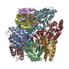
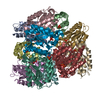
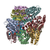
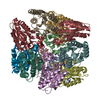
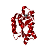
| Title | Crystal structure of type II dehydroquinase from Streptomyces coelicolor complexed with 2,3-anhydro-quinic acid | ||||||
 Components Components | 3-DEHYDROQUINATE DEHYDRATASE | ||||||
 Keywords Keywords | LYASE / SHIKIMATE PATHWAY / TETRAHEDRAL SYMMETRY | ||||||
| Function / homology |  Function and homology information Function and homology informationquinate catabolic process / 3-dehydroquinate dehydratase / 3-dehydroquinate dehydratase activity / chorismate biosynthetic process / aromatic amino acid biosynthetic process / amino acid biosynthetic process Similarity search - Function | ||||||
| Biological species |  STREPTOMYCES COELICOLOR (bacteria) STREPTOMYCES COELICOLOR (bacteria) | ||||||
| Method |  X-RAY DIFFRACTION / SYNCHROTRON / MOLECULAR REPLACEMENT / Resolution: 1.8 Å X-RAY DIFFRACTION / SYNCHROTRON / MOLECULAR REPLACEMENT / Resolution: 1.8 Å | ||||||
 Authors Authors | Roszak, A.W. / Robinson, D.A. / Krell, T. / Hunter, I.S. / Coggins, J.R. / Lapthorn, A.J. | ||||||
 Citation Citation | Journal: Structure / Year: 2002 Title: The Structure and Mechanism of the Type II Dehydroquinase from Streptomyces Coelicolor Authors: Roszak, A.W. / Robinson, D.A. / Krell, T. / Hunter, I.S. / Fredrickson, M. / Abell, C. / Coggins, J.R. / Lapthorn, A.J. | ||||||
| History |
|
- Structure visualization
| Structure viewer | Molecule: MolmilJmol/JSmol |
|---|
- Downloads & links
-Download
| PDBx/mmCIF format | 1gu1.cif.gz | 772.8 KB | Display | PDBx/mmCIF format |
|---|---|---|---|---|
| PDB format | pdb1gu1.ent.gz | 647.8 KB | Display | PDB format |
| PDBx/mmJSON format | 1gu1.json.gz | Tree view | PDBx/mmJSON format | |
| Others |  Other downloads Other downloads |
-Validation report
| Arichive directory | https://data.pdbj.org/pub/pdb/validation_reports/gu/1gu1ftp://data.pdbj.org/pub/pdb/validation_reports/gu/1gu1 | HTTPS FTP |
|---|
-Related structure data
| Related structure data |  1d0iC  1gtzC  1gu0C  2dhqS C: citing same article ( S: Starting model for refinement |
|---|---|
| Similar structure data |


-Links
 PDBj
PDBj

- Assembly
| Deposited unit | 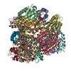
| ||||||||||||||||||||||||||||||||||||||||||||||||
|---|---|---|---|---|---|---|---|---|---|---|---|---|---|---|---|---|---|---|---|---|---|---|---|---|---|---|---|---|---|---|---|---|---|---|---|---|---|---|---|---|---|---|---|---|---|---|---|---|---|
| 1 |
| ||||||||||||||||||||||||||||||||||||||||||||||||
| Unit cell |
| ||||||||||||||||||||||||||||||||||||||||||||||||
| Noncrystallographic symmetry (NCS) | NCS oper:
|
-Components
-Protein , 1 types, 12 molecules ABCDEFGHIJKL
| #1: Protein | Mass: 16569.605 Da / Num. of mol.: 12 Source method: isolated from a genetically manipulated source Source: (gene. exp.) STREPTOMYCES COELICOLOR (bacteria) / Plasmid: PDHQ / Production host: |
|---|
-Non-polymers , 5 types, 2674 molecules 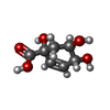


| #2: Chemical | ChemComp-FA1 /  Mass: 174.151 Da / Num. of mol.: 12 / Source method: obtained synthetically / Formula: C7H10O5 Mass: 174.151 Da / Num. of mol.: 12 / Source method: obtained synthetically / Formula: C7H10O5#3: Chemical | ChemComp-GOL /  Mass: 92.094 Da / Num. of mol.: 12 / Source method: obtained synthetically / Formula: C3H8O3 Mass: 92.094 Da / Num. of mol.: 12 / Source method: obtained synthetically / Formula: C3H8O3#4: Chemical | ChemComp-TLA /  Mass: 150.087 Da / Num. of mol.: 12 / Source method: obtained synthetically / Formula: C4H6O6 Mass: 150.087 Da / Num. of mol.: 12 / Source method: obtained synthetically / Formula: C4H6O6#5: Chemical | ChemComp-TRS /  Mass: 122.143 Da / Num. of mol.: 4 / Source method: obtained synthetically / Formula: C4H12NO3 / Comment: pH buffer*YM Mass: 122.143 Da / Num. of mol.: 4 / Source method: obtained synthetically / Formula: C4H12NO3 / Comment: pH buffer*YM#6: Water | ChemComp-HOH / | Mass: 18.015 Da / Num. of mol.: 2634 / Source method: isolated from a natural source / Formula: H2O |
|---|
-Experimental details
-Experiment
| Experiment | Method: X-RAY DIFFRACTION / Number of used crystals: 1 |
|---|
- Sample preparation
| Crystal | Density Matthews: 2.8 Å3/Da / Density % sol: 55.9 % |
|---|---|
| Crystal grow | pH: 8.5 Details: PEG 8000, SODIUM/POTASSIUM PHOSPHATE, TRIS BUFFER, pH 8.50 |
-Data collection
| Diffraction | Mean temperature: 100 K |
|---|---|
| Diffraction source | Source: SYNCHROTRON / Site: SRS  / Beamline: PX9.6 / Wavelength: 0.89 / Beamline: PX9.6 / Wavelength: 0.89 |
| Detector | Type: ADSC QUANTUM 4 / Detector: CCD / Date: Mar 15, 1998 |
| Radiation | Protocol: SINGLE WAVELENGTH / Monochromatic (M) / Laue (L): M / Scattering type: x-ray |
| Radiation wavelength | Wavelength: 0.89 Å / Relative weight: 1 |
| Reflection | Resolution: 1.8→70 Å / Num. obs: 648516 / % possible obs: 98.1 % / Observed criterion σ(I): 0 / Redundancy: 4 % / Rmerge(I) obs: 0.088 |
| Reflection shell | Resolution: 1.8→1.82 Å / Redundancy: 2.9 % / % possible all: 90.9 |
- Processing
| Software |
| ||||||||||||||||||||
|---|---|---|---|---|---|---|---|---|---|---|---|---|---|---|---|---|---|---|---|---|---|
| Refinement | Method to determine structure: MOLECULAR REPLACEMENT Starting model: PDB ENTRY 2DHQ Resolution: 1.8→22 Å / Cross valid method: THROUGHOUT / σ(F): 0 / Details: NONE
| ||||||||||||||||||||
| Refinement step | Cycle: LAST / Resolution: 1.8→22 Å
|
