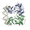
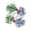
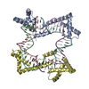
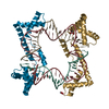
- PDB-5jgh: Crystal structure of the mitochondrial DNA packaging protein Abf2... -
+
Open data
ID or keywords:
Loading...
-
Basic information
Entry
Database: PDB / ID: 5jgh
Title
Crystal structure of the mitochondrial DNA packaging protein Abf2p in complex with DNA at 2.6 Angstrom resolution
Components
ARS-binding factor 2, mitochondrial
DNA (5'-D(*TP*TP*AP*TP*AP*TP*AP*AP*TP*AP*TP*AP*AP*AP*AP*TP*AP*AP*TP*AP*AP*A)-3')
DNA (5'-D(*TP*TP*TP*AP*TP*TP*AP*TP*TP*TP*TP*AP*TP*AP*TP*TP*AP*TP*AP*TP*AP*A)-3')
Keywords
DNA BINDING PROTEIN / DNA binding protein DNA packaging
Function / homology
Function and homology information
mitochondrial chromosome packaging / Apoptosis induced DNA fragmentation / Regulation of TLR by endogenous ligand / mitochondrion inheritance / Mitochondrial transcription initiation / Pyroptosis / mitochondrial chromosome / Mitochondrial protein degradation / DNA binding, bending / mitochondrial nucleoid ...mitochondrial chromosome packaging / Apoptosis induced DNA fragmentation / Regulation of TLR by endogenous ligand / mitochondrion inheritance / Mitochondrial transcription initiation / Pyroptosis / mitochondrial chromosome / Mitochondrial protein degradation / DNA binding, bending / mitochondrial nucleoid / Neutrophil degranulation / chromatin remodeling / mitochondrion / DNA binding / nucleus Similarity search - Function
: / HMG (high mobility group) box / HMG boxes A and B DNA-binding domains profile. / high mobility group / High mobility group box domain / High mobility group box domain superfamily Similarity search - Domain/homology
In the structure databanks used in Yorodumi, some data are registered as the other names, "COVID-19 virus" and "2019-nCoV". Here are the details of the virus and the list of structure data.
Jan 31, 2019. EMDB accession codes are about to change! (news from PDBe EMDB page)
EMDB accession codes are about to change! (news from PDBe EMDB page)
The allocation of 4 digits for EMDB accession codes will soon come to an end. Whilst these codes will remain in use, new EMDB accession codes will include an additional digit and will expand incrementally as the available range of codes is exhausted. The current 4-digit format prefixed with “EMD-” (i.e. EMD-XXXX) will advance to a 5-digit format (i.e. EMD-XXXXX), and so on. It is currently estimated that the 4-digit codes will be depleted around Spring 2019, at which point the 5-digit format will come into force.
The EM Navigator/Yorodumi systems omit the EMD- prefix.
Related info.:Q: What is EMD? / ID/Accession-code notation in Yorodumi/EM Navigator
Yorodumi is a browser for structure data from EMDB, PDB, SASBDB, etc.
This page is also the successor to EM Navigator detail page, and also detail information page/front-end page for Omokage search.
The word "yorodu" (or yorozu) is an old Japanese word meaning "ten thousand". "mi" (miru) is to see.
Related info.:EMDB / PDB / SASBDB / Comparison of 3 databanks / Yorodumi Search / Aug 31, 2016. New EM Navigator & Yorodumi / Yorodumi Papers / Jmol/JSmol / Function and homology information / Changes in new EM Navigator and Yorodumi
 Movie
Movie Controller

 Yorodumi
 Open data
 Basic information
 Components
 Keywords
 Function and homology information
 X-RAY DIFFRACTION /
  Authors
 Spain, 1items
  Citation
 Structure visualization
 Downloads & links
 Other downloads
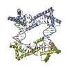

 PDBj
PDBj


 Assembly
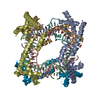


 Mass: 59.044 Da / Num. of mol.: 2 / Source method: obtained synthetically / Formula: C2H3O2
 Mass: 18.015 Da / Num. of mol.: 126 / Source method: isolated from a natural source / Formula: H2O
 Sample preparation
 / Beamline: ID29 / Wavelength: 0.97625 Å
 Processing