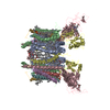
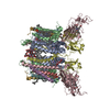
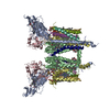
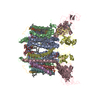
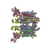
#1: Journal: Proc.Natl.Acad.Sci.USA / Year: 2003 Title: A Defined Protein-Detergent-Lipid Complex for Crystallization of Integral Membrane Proteins: The Cytochrome B6F Complex of Oxygenic Photosynthesis Authors: Zhang, H. / Kurisu, G. / Smith, J.L. / Cramer, W.A.
A: CYTOCHROME B6 B: SUBUNIT IV C: CYTOCHROME F D: RIESKE IRON-SULFUR PROTEIN E: PROTEIN PET L F: PROTEIN PET M G: PROTEIN PET G H: PROTEIN PET N N: CYTOCHROME B6 O: SUBUNIT IV P: CYTOCHROME F Q: RIESKE IRON-SULFUR PROTEIN R: PROTEIN PET L S: PROTEIN PET M T: PROTEIN PET G U: PROTEIN PET N hetero molecules
Monochromator: SI(111) / Protocol: SINGLE WAVELENGTH / Monochromatic (M) / Laue (L): M / Scattering type: x-ray
Radiation wavelength
Wavelength: 1.0332 Å / Relative weight: 1
Reflection
Resolution: 3→150 Å / Num. obs: 100622 / % possible obs: 99.8 % / Observed criterion σ(I): -2 / Redundancy: 7 % / Rmerge(I) obs: 0.068 / Net I/σ(I): 17.9
Reflection shell
Resolution: 3→3.11 Å / Rmerge(I) obs: 0.54 / Mean I/σ(I) obs: 2.2 / % possible all: 100
-
Processing
Software
Name
Version
Classification
HKL-2000
datacollection
HKL-2000
datareduction
MLPHARE
phasing
CNS
1.1
refinement
HKL-2000
datascaling
Refinement
Method to determine structure: MIR / Resolution: 3→48.16 Å / Data cutoff high absF: 2928724.46 / Data cutoff low absF: 0 / Isotropic thermal model: GROUP / Cross valid method: THROUGHOUT / σ(F): 0 / Stereochemistry target values: ENGH & HUBER Details: Twinning was treated during refinement and the twinning fraction set to 0.50.
In the structure databanks used in Yorodumi, some data are registered as the other names, "COVID-19 virus" and "2019-nCoV". Here are the details of the virus and the list of structure data.
Jan 31, 2019. EMDB accession codes are about to change! (news from PDBe EMDB page)
EMDB accession codes are about to change! (news from PDBe EMDB page)
The allocation of 4 digits for EMDB accession codes will soon come to an end. Whilst these codes will remain in use, new EMDB accession codes will include an additional digit and will expand incrementally as the available range of codes is exhausted. The current 4-digit format prefixed with “EMD-” (i.e. EMD-XXXX) will advance to a 5-digit format (i.e. EMD-XXXXX), and so on. It is currently estimated that the 4-digit codes will be depleted around Spring 2019, at which point the 5-digit format will come into force.
The EM Navigator/Yorodumi systems omit the EMD- prefix.
Related info.:Q: What is EMD? / ID/Accession-code notation in Yorodumi/EM Navigator
Yorodumi is a browser for structure data from EMDB, PDB, SASBDB, etc.
This page is also the successor to EM Navigator detail page, and also detail information page/front-end page for Omokage search.
The word "yorodu" (or yorozu) is an old Japanese word meaning "ten thousand". "mi" (miru) is to see.
Related info.:EMDB / PDB / SASBDB / Comparison of 3 databanks / Yorodumi Search / Aug 31, 2016. New EM Navigator & Yorodumi / Yorodumi Papers / Jmol/JSmol / Function and homology information / Changes in new EM Navigator and Yorodumi
 Movie
Movie Controller


 Open data
 Basic information
 Components
 Keywords
 Function and homology information
 Mastigocladus laminosus (bacteria)
 X-RAY DIFFRACTION /
  Authors
 Citation
 Structure visualization
 Downloads & links
 Other downloads
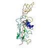


 PDBj
PDBj


 Assembly


 Mass: 616.487 Da / Num. of mol.: 8 / Source method: obtained synthetically / Formula: C34H32FeN4O4
 Mass: 418.566 Da / Num. of mol.: 2 / Source method: obtained synthetically / Formula: C25H38O5
 Mass: 749.201 Da / Num. of mol.: 2 / Source method: obtained synthetically / Formula: C53H80O2
 Mass: 801.148 Da / Num. of mol.: 4 / Source method: obtained synthetically / Formula: C45H87NO8P / Comment: phospholipid*YM
 Mass: 893.489 Da / Num. of mol.: 2 / Source method: obtained synthetically / Formula: C55H72MgN4O5
 Mass: 175.820 Da / Num. of mol.: 2 / Source method: obtained synthetically / Formula: Fe2S2
 Mass: 536.873 Da / Num. of mol.: 2 / Source method: obtained synthetically / Formula: C40H56
 Sample preparation
 / Beamline: 19-ID / Wavelength: 1.0332
  Processing