 Movie
Movie Controller


+ Open data

- Basic information
| Entry | Database: PDB / ID: 1ci3 | |||||||||
|---|---|---|---|---|---|---|---|---|---|---|
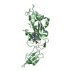
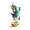
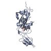
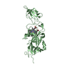
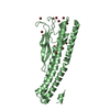
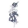
| Title | CYTOCHROME F FROM THE B6F COMPLEX OF PHORMIDIUM LAMINOSUM | |||||||||
 Components Components | PROTEIN (CYTOCHROME F) | |||||||||
 Keywords Keywords | ELECTRON TRANSPORT / ELECTRON TRANSFER PROTEIN / COMPLEX SUBUNIT | |||||||||
| Function / homology |  Function and homology information Function and homology informationplasma membrane-derived thylakoid membrane / photosynthesis / electron transfer activity / iron ion binding / heme binding Similarity search - Function | |||||||||
| Biological species |  Phormidium laminosum (bacteria) Phormidium laminosum (bacteria) | |||||||||
| Method |  X-RAY DIFFRACTION / MOLECULAR REPLACEMENT / Resolution: 1.9 Å X-RAY DIFFRACTION / MOLECULAR REPLACEMENT / Resolution: 1.9 Å | |||||||||
 Authors Authors | Carrell, C.J. / Schlarb, B.G. / Howe, C.J. / Bendall, D.S. / Cramer, W.A. / Smith, J.L. | |||||||||
 Citation Citation | Journal: Biochemistry / Year: 1999 Title: Structure of the soluble domain of cytochrome f from the cyanobacterium Phormidium laminosum. Authors: Carrell, C.J. / Schlarb, B.G. / Bendall, D.S. / Howe, C.J. / Cramer, W.A. / Smith, J.L. #1: Journal: Protein Sci. / Year: 1996Title: The Heme Redox Center of Chloroplast Cytochrome F is Linked to a Buried Five- Water Chain Authors: Martinez, S.E. / Huang, D. / Ponomarev, M. / Cramer, W.A. / Smith, J.L. | |||||||||
| History |
|
- Structure visualization
| Structure viewer | Molecule: MolmilJmol/JSmol |
|---|
- Downloads & links
-Download
| PDBx/mmCIF format | 1ci3.cif.gz | 68.9 KB | Display | PDBx/mmCIF format |
|---|---|---|---|---|
| PDB format | pdb1ci3.ent.gz | 49.8 KB | Display | PDB format |
| PDBx/mmJSON format | 1ci3.json.gz | Tree view | PDBx/mmJSON format | |
| Others |  Other downloads Other downloads |
-Validation report
| Arichive directory | https://data.pdbj.org/pub/pdb/validation_reports/ci/1ci3ftp://data.pdbj.org/pub/pdb/validation_reports/ci/1ci3 | HTTPS FTP |
|---|
-Related structure data
| Related structure data |  1hczS S: Starting model for refinement |
|---|---|
| Similar structure data |


-Links
 PDBj
PDBj


- Assembly
| Deposited unit | 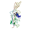
| ||||||||
|---|---|---|---|---|---|---|---|---|---|
| 1 |
| ||||||||
| 2 | x 6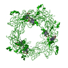
| ||||||||
| 3 | 
| ||||||||
| Unit cell |
|
-Components
| #1: Protein | Mass: 26462.555 Da / Num. of mol.: 1 / Fragment: SOLUBLE EXTRINSIC FRAGMENT Source method: isolated from a genetically manipulated source Details: THIOETHER LINK BETWEEN CYS 21 AND HEME 254; COORDINATION OF HEME IRON BY HIS 25 AND N-TERMINAL AMINO GROUP Source: (gene. exp.) Phormidium laminosum (bacteria) / Cellular location: MEMBRANE-CYTOPLASM INTERFACE / Gene: PETA / Plasmid: PUC19CF / Species (production host): Escherichia coliProduction host: Strain (production host): W3110 / References: UniProt: P95522 | ||||||
|---|---|---|---|---|---|---|---|
| #2: Chemical |   Mass: 65.409 Da / Num. of mol.: 2 / Source method: obtained synthetically / Formula: Zn Mass: 65.409 Da / Num. of mol.: 2 / Source method: obtained synthetically / Formula: Zn#3: Chemical | ChemComp-HEC / |   Mass: 618.503 Da / Num. of mol.: 1 / Source method: obtained synthetically / Formula: C34H34FeN4O4 Mass: 618.503 Da / Num. of mol.: 1 / Source method: obtained synthetically / Formula: C34H34FeN4O4#4: Water | ChemComp-HOH / |  Mass: 18.015 Da / Num. of mol.: 250 / Source method: isolated from a natural source / Formula: H2O Mass: 18.015 Da / Num. of mol.: 250 / Source method: isolated from a natural source / Formula: H2OHas protein modification | Y | |
-Experimental details
-Experiment
| Experiment | Method: X-RAY DIFFRACTION / Number of used crystals: 1 |
|---|
- Sample preparation
| Crystal | Density Matthews: 3.1 Å3/Da / Density % sol: 61.15 % | ||||||||||||||||||||||||||||||||||||||||||
|---|---|---|---|---|---|---|---|---|---|---|---|---|---|---|---|---|---|---|---|---|---|---|---|---|---|---|---|---|---|---|---|---|---|---|---|---|---|---|---|---|---|---|---|
| Crystal grow | pH: 6.5 / Details: pH 6.50 | ||||||||||||||||||||||||||||||||||||||||||
| Crystal | *PLUS Density % sol: 60 % | ||||||||||||||||||||||||||||||||||||||||||
| Crystal grow | *PLUS pH: 7.5 / Method: vapor diffusion, sitting drop | ||||||||||||||||||||||||||||||||||||||||||
| Components of the solutions | *PLUS
|
-Data collection
| Diffraction | Mean temperature: 110 K |
|---|---|
| Diffraction source | Source: ROTATING ANODE / Type: RIGAKU RU200 / Wavelength: 1.5418 |
| Detector | Type: RIGAKU RAXIS IV / Detector: IMAGE PLATE / Date: Feb 1, 1999 |
| Radiation | Protocol: SINGLE WAVELENGTH / Monochromatic (M) / Laue (L): M / Scattering type: x-ray |
| Radiation wavelength | Wavelength: 1.5418 Å / Relative weight: 1 |
| Reflection | Resolution: 1.9→40 Å / Num. obs: 26071 / % possible obs: 97.9 % / Redundancy: 4.8 % / Rsym value: 3.2 / Net I/σ(I): 42 |
| Reflection shell | Resolution: 1.9→1.97 Å / Redundancy: 2.3 % / Mean I/σ(I) obs: 8.5 / Rsym value: 12.8 / % possible all: 83.8 |
| Reflection | *PLUS Highest resolution: 1.9 Å / Lowest resolution: 40 Å / % possible obs: 97.9 % / Redundancy: 4.8 % / Num. measured all: 126348 / Rmerge(I) obs: 0.032 |
| Reflection shell | *PLUS Highest resolution: 1.9 Å / Lowest resolution: 1.97 Å / % possible obs: 83.8 % / Redundancy: 2.3 % / Rmerge(I) obs: 0.128 / Mean I/σ(I) obs: 8.5 |
- Processing
| Software |
| ||||||||||||||||||||||||||||||||||||||||||||||||||||||||||||||||||||||||||||||||
|---|---|---|---|---|---|---|---|---|---|---|---|---|---|---|---|---|---|---|---|---|---|---|---|---|---|---|---|---|---|---|---|---|---|---|---|---|---|---|---|---|---|---|---|---|---|---|---|---|---|---|---|---|---|---|---|---|---|---|---|---|---|---|---|---|---|---|---|---|---|---|---|---|---|---|---|---|---|---|---|---|---|
| Refinement | Method to determine structure: MOLECULAR REPLACEMENT Starting model: PDB ENTRY 1HCZ Resolution: 1.9→30 Å / Data cutoff high rms absF: 10000 / Isotropic thermal model: RESTRAINED / Cross valid method: THROUGHOUT / σ(F): 0
| ||||||||||||||||||||||||||||||||||||||||||||||||||||||||||||||||||||||||||||||||
| Solvent computation | Solvent model: FLAT MODEL / Bsol: 120 Å2 / ksol: 1 e/Å3 | ||||||||||||||||||||||||||||||||||||||||||||||||||||||||||||||||||||||||||||||||
| Displacement parameters |
| ||||||||||||||||||||||||||||||||||||||||||||||||||||||||||||||||||||||||||||||||
| Refine analyze |
| ||||||||||||||||||||||||||||||||||||||||||||||||||||||||||||||||||||||||||||||||
| Refinement step | Cycle: LAST / Resolution: 1.9→30 Å
| ||||||||||||||||||||||||||||||||||||||||||||||||||||||||||||||||||||||||||||||||
| Refine LS restraints |
| ||||||||||||||||||||||||||||||||||||||||||||||||||||||||||||||||||||||||||||||||
| LS refinement shell | Resolution: 1.9→1.92 Å / Total num. of bins used: 39
| ||||||||||||||||||||||||||||||||||||||||||||||||||||||||||||||||||||||||||||||||
| Software | *PLUS Name: CNS / Version: 0.5 / Classification: refinement | ||||||||||||||||||||||||||||||||||||||||||||||||||||||||||||||||||||||||||||||||
| Refinement | *PLUS Highest resolution: 1.9 Å / Lowest resolution: 30 Å / Rfactor obs: 0.203 | ||||||||||||||||||||||||||||||||||||||||||||||||||||||||||||||||||||||||||||||||
| Solvent computation | *PLUS | ||||||||||||||||||||||||||||||||||||||||||||||||||||||||||||||||||||||||||||||||
| Displacement parameters | *PLUS | ||||||||||||||||||||||||||||||||||||||||||||||||||||||||||||||||||||||||||||||||
| Refine LS restraints | *PLUS
| ||||||||||||||||||||||||||||||||||||||||||||||||||||||||||||||||||||||||||||||||
| LS refinement shell | *PLUS Rfactor Rfree: 0.288 / Rfactor Rwork: 0.233 |
