 Movie
Movie Controller


+ Open data

- Basic information
| Entry | Database: PDB / ID: 1e2w | ||||||
|---|---|---|---|---|---|---|---|
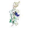
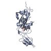
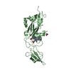
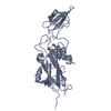
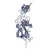
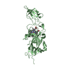
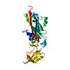
| Title | N168F mutant of cytochrome f from Chlamydomonas reinhardtii | ||||||
 Components Components | CYTOCHROME F | ||||||
 Keywords Keywords | ELECTRON TRANSPORT PROTEINS / INTERNAL WATER CHAIN / PHOTOSYNTHETIC FUNCTION IMPAIRED | ||||||
| Function / homology |  Function and homology information Function and homology informationchloroplast thylakoid membrane / photosynthesis / electron transfer activity / iron ion binding / heme binding Similarity search - Function | ||||||
| Biological species |   CHLAMYDOMONAS REINHARDTII (plant) CHLAMYDOMONAS REINHARDTII (plant) | ||||||
| Method |  X-RAY DIFFRACTION / SYNCHROTRON / MOLECULAR REPLACEMENT / Resolution: 1.6 Å X-RAY DIFFRACTION / SYNCHROTRON / MOLECULAR REPLACEMENT / Resolution: 1.6 Å | ||||||
 Authors Authors | Sainz, G. / Carrell, C.J. / Ponamarev, M.V. / Soriano, G.M. / Cramer, W.A. / Smith, J.L. | ||||||
 Citation Citation | Journal: Biochemistry / Year: 2000 Title: Interruption of the Internal Water Chain of Cytochrome F Impairs Photosynthetic Function Authors: Sainz, G. / Carrell, C.J. / Ponamarev, M.V. / Soriano, G.M. / Cramer, W.A. / Smith, J.L. | ||||||
| History |
|
- Structure visualization
| Structure viewer | Molecule: MolmilJmol/JSmol |
|---|
- Downloads & links
-Download
| PDBx/mmCIF format | 1e2w.cif.gz | 127.6 KB | Display | PDBx/mmCIF format |
|---|---|---|---|---|
| PDB format | pdb1e2w.ent.gz | 99.1 KB | Display | PDB format |
| PDBx/mmJSON format | 1e2w.json.gz | Tree view | PDBx/mmJSON format | |
| Others |  Other downloads Other downloads |
-Validation report
| Arichive directory | https://data.pdbj.org/pub/pdb/validation_reports/e2/1e2wftp://data.pdbj.org/pub/pdb/validation_reports/e2/1e2w | HTTPS FTP |
|---|
-Related structure data
| Related structure data |  1e2vSC  1e2zC  1ewhC S: Starting model for refinement C: citing same article ( |
|---|---|
| Similar structure data |


-Links
 PDBj
PDBj


- Assembly
| Deposited unit | 
| ||||||||
|---|---|---|---|---|---|---|---|---|---|
| 1 | 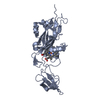
| ||||||||
| 2 | 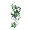
| ||||||||
| Unit cell |
|
-Components
| #1: Protein | Mass: 27215.033 Da / Num. of mol.: 2 / Mutation: YES Source method: isolated from a genetically manipulated source Source: (gene. exp.) CHLAMYDOMONAS REINHARDTII (plant) / Gene: PETA / Plasmid: PUCPF2 / Gene (production host): CCMABCDEFGH / Production host:  #2: Chemical |   Mass: 618.503 Da / Num. of mol.: 2 / Source method: obtained synthetically / Formula: C34H34FeN4O4 Mass: 618.503 Da / Num. of mol.: 2 / Source method: obtained synthetically / Formula: C34H34FeN4O4#3: Water | ChemComp-HOH / |  Mass: 18.015 Da / Num. of mol.: 757 / Source method: isolated from a natural source / Formula: H2O Mass: 18.015 Da / Num. of mol.: 757 / Source method: isolated from a natural source / Formula: H2OCompound details | CHAIN A, B, C ENGINEERED MUTATION ASN168PHE. ONLY THE SOLUBLE DOMAIN OF THE MATURE MEMBRANE PROTEIN ...CHAIN A, B, C ENGINEERED | Has protein modification | Y | |
|---|
-Experimental details
-Experiment
| Experiment | Method: X-RAY DIFFRACTION / Number of used crystals: 1 |
|---|
- Sample preparation
| Crystal | Density Matthews: 2.62 Å3/Da / Density % sol: 52.6 % | |||||||||||||||||||||||||||||||||||
|---|---|---|---|---|---|---|---|---|---|---|---|---|---|---|---|---|---|---|---|---|---|---|---|---|---|---|---|---|---|---|---|---|---|---|---|---|
| Crystal grow | pH: 6.7 Details: THE PROTEIN WAS BUFFERED IN 10 MM NA2HPO4/NAH2PO4, PH 7.5, 1 MM DTT, THE RESERVOIR CONTAINED 100 MM MES, PH 6.7, 200 MM AMMONIUM FORMATE, 14% GLYCEROL, 17% PEG-3350. | |||||||||||||||||||||||||||||||||||
| Crystal grow | *PLUS pH: 7.5 / Method: vapor diffusion | |||||||||||||||||||||||||||||||||||
| Components of the solutions | *PLUS
|
-Data collection
| Diffraction | Mean temperature: 100 K |
|---|---|
| Diffraction source | Source: SYNCHROTRON / Site: APS  / Beamline: 19-ID / Wavelength: 0.6526 / Beamline: 19-ID / Wavelength: 0.6526 |
| Detector | Type: CUSTOM-MADE / Detector: CCD / Date: Apr 15, 1999 / Details: MIRROR |
| Radiation | Protocol: SINGLE WAVELENGTH / Monochromatic (M) / Laue (L): M / Scattering type: x-ray |
| Radiation wavelength | Wavelength: 0.6526 Å / Relative weight: 1 |
| Reflection | Resolution: 1.6→27.84 Å / Num. obs: 73580 / % possible obs: 99.6 % / Redundancy: 3.8 % / Biso Wilson estimate: 18.6 Å2 / Rmerge(I) obs: 0.046 / Rsym value: 0.046 / Net I/σ(I): 28.6 |
| Reflection shell | Resolution: 1.6→1.66 Å / Redundancy: 3.5 % / Rmerge(I) obs: 0.378 / Mean I/σ(I) obs: 3.54 / Rsym value: 0.378 / % possible all: 100 |
| Reflection | *PLUS Num. measured all: 277890 |
| Reflection shell | *PLUS % possible obs: 100 % |
- Processing
| Software |
| ||||||||||||||||||||||||||||||||||||||||||||||||||||||||||||||||||||||||||||||||
|---|---|---|---|---|---|---|---|---|---|---|---|---|---|---|---|---|---|---|---|---|---|---|---|---|---|---|---|---|---|---|---|---|---|---|---|---|---|---|---|---|---|---|---|---|---|---|---|---|---|---|---|---|---|---|---|---|---|---|---|---|---|---|---|---|---|---|---|---|---|---|---|---|---|---|---|---|---|---|---|---|---|
| Refinement | Method to determine structure: MOLECULAR REPLACEMENT Starting model: 1E2V Resolution: 1.6→27.41 Å / Rfactor Rfree error: 0.004 / Data cutoff high absF: 1175031.81 / Isotropic thermal model: RESTRAINED / Cross valid method: THROUGHOUT / σ(F): 0 Details: THE LOOP 186-190 IS BADLY DEFINED IN THE ELECTRON DENSITY. ONLY THE MAIN CHAIN OF THE ARG 251 WAS SEEN IN THE ELCTRON DENSITY MAP FOR THE B CHAIN , SO AN ALA 251 WAS USED
| ||||||||||||||||||||||||||||||||||||||||||||||||||||||||||||||||||||||||||||||||
| Solvent computation | Solvent model: FLAT MODEL / Bsol: 54.4172 Å2 / ksol: 0.359825 e/Å3 | ||||||||||||||||||||||||||||||||||||||||||||||||||||||||||||||||||||||||||||||||
| Displacement parameters | Biso mean: 26.1 Å2
| ||||||||||||||||||||||||||||||||||||||||||||||||||||||||||||||||||||||||||||||||
| Refine analyze |
| ||||||||||||||||||||||||||||||||||||||||||||||||||||||||||||||||||||||||||||||||
| Refinement step | Cycle: LAST / Resolution: 1.6→27.41 Å
| ||||||||||||||||||||||||||||||||||||||||||||||||||||||||||||||||||||||||||||||||
| Refine LS restraints |
| ||||||||||||||||||||||||||||||||||||||||||||||||||||||||||||||||||||||||||||||||
| LS refinement shell | Resolution: 1.6→1.7 Å / Rfactor Rfree error: 0.012 / Total num. of bins used: 6
| ||||||||||||||||||||||||||||||||||||||||||||||||||||||||||||||||||||||||||||||||
| Xplor file |
| ||||||||||||||||||||||||||||||||||||||||||||||||||||||||||||||||||||||||||||||||
| Software | *PLUS Name: CNS / Version: 1 / Classification: refinement | ||||||||||||||||||||||||||||||||||||||||||||||||||||||||||||||||||||||||||||||||
| Refine LS restraints | *PLUS
|
