 Movie
Movie Controller


+ Open data

- Basic information
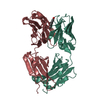
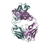
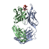
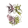
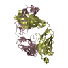
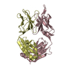
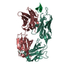
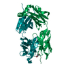
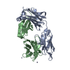
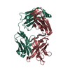
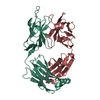
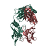
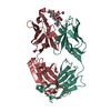
| Entry | Database: PDB / ID: 1axs | ||||||
|---|---|---|---|---|---|---|---|
| Title | MATURE OXY-COPE CATALYTIC ANTIBODY WITH HAPTEN | ||||||
 Components Components | (OXY-COPE CATALYTIC ANTIBODY) x 2 | ||||||
 Keywords Keywords | CATALYTIC ANTIBODY / OXY-COPE / FAB FRAGMENT | ||||||
| Function / homology |  Function and homology information Function and homology informationcomplement-dependent cytotoxicity / antibody-dependent cellular cytotoxicity / Fc-gamma receptor I complex binding / immunoglobulin complex, circulating / Classical antibody-mediated complement activation / immunoglobulin receptor binding / Initial triggering of complement / IgG immunoglobulin complex / FCGR activation / complement activation, classical pathway ...complement-dependent cytotoxicity / antibody-dependent cellular cytotoxicity / Fc-gamma receptor I complex binding / immunoglobulin complex, circulating / Classical antibody-mediated complement activation / immunoglobulin receptor binding / Initial triggering of complement / IgG immunoglobulin complex / FCGR activation / complement activation, classical pathway / Role of phospholipids in phagocytosis / immunoglobulin complex / antigen binding / FCGR3A-mediated IL10 synthesis / Regulation of Complement cascade / B cell receptor signaling pathway / FCGR3A-mediated phagocytosis / Regulation of actin dynamics for phagocytic cup formation / antibacterial humoral response / Interleukin-4 and Interleukin-13 signaling / blood microparticle / adaptive immune response / : / extracellular exosome / extracellular region / plasma membrane Similarity search - Function | ||||||
| Biological species |  Homo sapiens (human) Homo sapiens (human) | ||||||
| Method |  X-RAY DIFFRACTION / SYNCHROTRON / MOLECULAR REPLACEMENT / Resolution: 2.6 Å X-RAY DIFFRACTION / SYNCHROTRON / MOLECULAR REPLACEMENT / Resolution: 2.6 Å | ||||||
 Authors Authors | Mundorff, E.C. / Ulrich, H.D. / Stevens, R.C. | ||||||
 Citation Citation | Journal: Nature / Year: 1997 Title: The interplay between binding energy and catalysis in the evolution of a catalytic antibody. Authors: Ulrich, H.D. / Mundorff, E. / Santarsiero, B.D. / Driggers, E.M. / Stevens, R.C. / Schultz, P.G. | ||||||
| History |
|
- Structure visualization
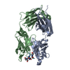
| Structure viewer | Molecule: MolmilJmol/JSmol |
|---|
- Downloads & links
-Download
| PDBx/mmCIF format | 1axs.cif.gz | 185.6 KB | Display | PDBx/mmCIF format |
|---|---|---|---|---|
| PDB format | pdb1axs.ent.gz | 146.2 KB | Display | PDB format |
| PDBx/mmJSON format | 1axs.json.gz | Tree view | PDBx/mmJSON format | |
| Others |  Other downloads Other downloads |
-Validation report
| Arichive directory | https://data.pdbj.org/pub/pdb/validation_reports/ax/1axsftp://data.pdbj.org/pub/pdb/validation_reports/ax/1axs | HTTPS FTP |
|---|
-Related structure data
| Related structure data | 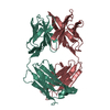 6fabS S: Starting model for refinement |
|---|---|
| Similar structure data |


-Links
 PDBj
PDBj


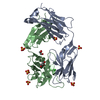
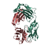
- Assembly
| Deposited unit | 
| |||||||||||||||||||||||||||||||||||||||
|---|---|---|---|---|---|---|---|---|---|---|---|---|---|---|---|---|---|---|---|---|---|---|---|---|---|---|---|---|---|---|---|---|---|---|---|---|---|---|---|---|
| 1 | 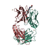
| |||||||||||||||||||||||||||||||||||||||
| 2 | 
| |||||||||||||||||||||||||||||||||||||||
| Unit cell |
| |||||||||||||||||||||||||||||||||||||||
| Noncrystallographic symmetry (NCS) | NCS domain:
NCS oper:
|
-Components
| #1: Antibody | Mass: 23529.176 Da / Num. of mol.: 2 / Fragment: FAB FRAGMENT Source method: isolated from a genetically manipulated source Details: CHIMERIC FAB FRAGMENT / Source: (gene. exp.) Homo sapiens (human) / Strain: SWISS WEBSTERDescription: THE PROTEIN WAS PRODUCED AS CHIMERIC FAB FRAGMENT. THE CONSTANT DOMAINS ARE HUMAN, THE VARIABLE DOMAINS ARE MURINE. PRODUCTION BY EXPRESSION FROM PHOA PROMOTER Cell: B LYMPHOCYTE / Cell line: AZ-28 HYBRIDOMA / Fragment: CHAIN L, A, 108 - 211, CHAIN H, B, 114 - 214 / Organ: SPLEEN / Plasmid: PAZ-28 / Cellular location (production host): PERIPLASM / Production host:  #2: Antibody | Mass: 23610.342 Da / Num. of mol.: 2 / Fragment: FAB FRAGMENT Source method: isolated from a genetically manipulated source Details: CHIMERIC FAB FRAGMENT / Source: (gene. exp.) Homo sapiens (human) / Strain: SWISS WEBSTERDescription: THE PROTEIN WAS PRODUCED AS CHIMERIC FAB FRAGMENT. THE CONSTANT DOMAINS ARE HUMAN, THE VARIABLE DOMAINS ARE MURINE. PRODUCTION BY EXPRESSION FROM PHOA PROMOTER Cell: B LYMPHOCYTE / Cell line: AZ-28 HYBRIDOMA / Fragment: CHAIN L, A, 108 - 211, CHAIN H, B, 114 - 214 / Organ: SPLEEN / Plasmid: PAZ-28 / Cellular location (production host): PERIPLASM / Production host: #3: Chemical | ChemComp-CD /   Mass: 112.411 Da / Num. of mol.: 8 / Source method: obtained synthetically / Formula: Cd Mass: 112.411 Da / Num. of mol.: 8 / Source method: obtained synthetically / Formula: Cd#4: Chemical | 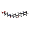  Mass: 381.465 Da / Num. of mol.: 2 / Source method: obtained synthetically / Formula: C23H27NO4 Mass: 381.465 Da / Num. of mol.: 2 / Source method: obtained synthetically / Formula: C23H27NO4#5: Water | ChemComp-HOH / |  Mass: 18.015 Da / Num. of mol.: 185 / Source method: isolated from a natural source / Formula: H2O Mass: 18.015 Da / Num. of mol.: 185 / Source method: isolated from a natural source / Formula: H2OHas protein modification | Y | |
|---|
-Experimental details
-Experiment
| Experiment | Method: X-RAY DIFFRACTION / Number of used crystals: 1 |
|---|
- Sample preparation
| Crystal | Density Matthews: 2.3 Å3/Da / Density % sol: 40 % | ||||||||||||||||||||||||||||||||||||
|---|---|---|---|---|---|---|---|---|---|---|---|---|---|---|---|---|---|---|---|---|---|---|---|---|---|---|---|---|---|---|---|---|---|---|---|---|---|
| Crystal grow | pH: 4.6 Details: PROTEIN WAS CRYSTALLIZED FROM 25%PEG 1000, 0.1 M SODIUM ACETATE, PH 4.6, 0.3 M CDCL2 AND 0.1 M AMMONIUM SULFATE IN THE PRESENCE OF 2MM HAPTEN. | ||||||||||||||||||||||||||||||||||||
| Crystal | *PLUS | ||||||||||||||||||||||||||||||||||||
| Crystal grow | *PLUS Method: vapor diffusion, hanging drop | ||||||||||||||||||||||||||||||||||||
| Components of the solutions | *PLUS
|
-Data collection
| Diffraction | Mean temperature: 94 K |
|---|---|
| Diffraction source | Source: SYNCHROTRON / Site: SSRL  / Beamline: BL7-1 / Wavelength: 1.08 / Beamline: BL7-1 / Wavelength: 1.08 |
| Detector | Type: MARRESEARCH / Detector: IMAGE PLATE / Date: Jan 1, 1997 / Details: MIRRORS |
| Radiation | Monochromator: SI(111) / Monochromatic (M) / Laue (L): M / Scattering type: x-ray |
| Radiation wavelength | Wavelength: 1.08 Å / Relative weight: 1 |
| Reflection | Resolution: 2.6→50 Å / Num. obs: 24768 / % possible obs: 90.9 % / Observed criterion σ(I): 2 / Redundancy: 4 % / Biso Wilson estimate: 34 Å2 / Rmerge(I) obs: 0.06 / Rsym value: 0.06 / Net I/σ(I): 16 |
| Reflection shell | Resolution: 2.6→2.69 Å / Redundancy: 3.5 % / Rmerge(I) obs: 0.142 / Mean I/σ(I) obs: 6 / Rsym value: 0.142 / % possible all: 87.5 |
- Processing
| Software |
| ||||||||||||||||||||||||||||||||||||||||||||||||||||||||||||||||||||||||||||||||
|---|---|---|---|---|---|---|---|---|---|---|---|---|---|---|---|---|---|---|---|---|---|---|---|---|---|---|---|---|---|---|---|---|---|---|---|---|---|---|---|---|---|---|---|---|---|---|---|---|---|---|---|---|---|---|---|---|---|---|---|---|---|---|---|---|---|---|---|---|---|---|---|---|---|---|---|---|---|---|---|---|---|
| Refinement | Method to determine structure: MOLECULAR REPLACEMENT Starting model: PDB ENTRY 6FAB Resolution: 2.6→20 Å / Rfactor Rfree error: 0.0062 / Data cutoff high absF: 1000000 / Data cutoff low absF: 0.0001 / Isotropic thermal model: RESTRAINED / Cross valid method: THROUGHOUT / σ(F): 2
| ||||||||||||||||||||||||||||||||||||||||||||||||||||||||||||||||||||||||||||||||
| Displacement parameters | Biso mean: 36.8 Å2 | ||||||||||||||||||||||||||||||||||||||||||||||||||||||||||||||||||||||||||||||||
| Refine analyze |
| ||||||||||||||||||||||||||||||||||||||||||||||||||||||||||||||||||||||||||||||||
| Refinement step | Cycle: LAST / Resolution: 2.6→20 Å
| ||||||||||||||||||||||||||||||||||||||||||||||||||||||||||||||||||||||||||||||||
| Refine LS restraints |
| ||||||||||||||||||||||||||||||||||||||||||||||||||||||||||||||||||||||||||||||||
| Refine LS restraints NCS | Refine-ID: X-RAY DIFFRACTION / Weight Biso : 2 / Weight position: 300
| ||||||||||||||||||||||||||||||||||||||||||||||||||||||||||||||||||||||||||||||||
| LS refinement shell | Resolution: 2.6→2.72 Å / Rfactor Rfree error: 0.022 / Total num. of bins used: 8
| ||||||||||||||||||||||||||||||||||||||||||||||||||||||||||||||||||||||||||||||||
| Xplor file |
| ||||||||||||||||||||||||||||||||||||||||||||||||||||||||||||||||||||||||||||||||
| Software | *PLUS Name: X-PLOR / Version: 3.851 / Classification: refinement | ||||||||||||||||||||||||||||||||||||||||||||||||||||||||||||||||||||||||||||||||
| Refinement | *PLUS Rfactor obs: 0.2 / Rfactor Rwork: 0.2 | ||||||||||||||||||||||||||||||||||||||||||||||||||||||||||||||||||||||||||||||||
| Solvent computation | *PLUS | ||||||||||||||||||||||||||||||||||||||||||||||||||||||||||||||||||||||||||||||||
| Displacement parameters | *PLUS | ||||||||||||||||||||||||||||||||||||||||||||||||||||||||||||||||||||||||||||||||
| Refine LS restraints | *PLUS
|
