ムービー
ムービー コントローラー


+ データを開く

- 基本情報
基本情報
| 登録情報 | データベース: EMDB / ID: EMD-1121 | |||||||||
|---|---|---|---|---|---|---|---|---|---|---|
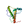
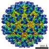
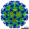
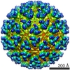
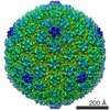
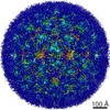
| タイトル | Mapping the structure and function of the E1 and E2 glycoproteins in alphaviruses. | |||||||||
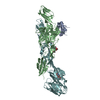
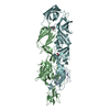
 マップデータ マップデータ | center of virus is where z=0 | |||||||||
 試料 試料 |
| |||||||||
| 機能・相同性 |  機能・相同性情報 機能・相同性情報icosahedral viral capsid, spike / togavirin / T=4 icosahedral viral capsid / host cell endoplasmic reticulum / ubiquitin-like protein ligase binding / channel activity / monoatomic ion transmembrane transport / clathrin-dependent endocytosis of virus by host cell / symbiont-mediated suppression of host toll-like receptor signaling pathway / membrane fusion ...icosahedral viral capsid, spike / togavirin / T=4 icosahedral viral capsid / host cell endoplasmic reticulum / ubiquitin-like protein ligase binding / channel activity / monoatomic ion transmembrane transport / clathrin-dependent endocytosis of virus by host cell / symbiont-mediated suppression of host toll-like receptor signaling pathway / membrane fusion / host cell Golgi apparatus / entry receptor-mediated virion attachment to host cell / serine-type endopeptidase activity / viral translational frameshifting / fusion of virus membrane with host endosome membrane / viral envelope / symbiont entry into host cell / virion attachment to host cell / host cell plasma membrane / host cell nucleus / virion membrane / structural molecule activity / proteolysis / RNA binding / identical protein binding 類似検索 - 分子機能 | |||||||||
| 生物種 |  Sindbis virus (シンドビスウイルス) Sindbis virus (シンドビスウイルス) | |||||||||
| 手法 | 単粒子再構成法 / クライオ電子顕微鏡法 / ネガティブ染色法 / 解像度: 9.0 Å | |||||||||
 データ登録者 データ登録者 | Mukhopadhyay S / Zhang W / Gabler S / Chipman PR / Strauss EG / Strauss JH / Baker TS / Kuhn RJ / Rossmann MG | |||||||||
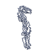
 引用 引用 | ジャーナル: Structure / 年: 2006 タイトル: Mapping the structure and function of the E1 and E2 glycoproteins in alphaviruses. 著者: Suchetana Mukhopadhyay / Wei Zhang / Stefan Gabler / Paul R Chipman / Ellen G Strauss / James H Strauss / Timothy S Baker / Richard J Kuhn / Michael G Rossmann /  要旨: The 9 A resolution cryo-electron microscopy map of Sindbis virus presented here provides structural information on the polypeptide topology of the E2 protein, on the interactions between the E1 and ...The 9 A resolution cryo-electron microscopy map of Sindbis virus presented here provides structural information on the polypeptide topology of the E2 protein, on the interactions between the E1 and E2 glycoproteins in the formation of a heterodimer, on the difference in conformation of the two types of trimeric spikes, on the interaction between the transmembrane helices of the E1 and E2 proteins, and on the conformational changes that occur when fusing with a host cell. The positions of various markers on the E2 protein established the approximate topology of the E2 structure. The largest conformational differences between the icosahedral surface spikes at icosahedral 3-fold and quasi-3-fold positions are associated with the monomers closest to the 5-fold axes. The long E2 monomers, containing the cell receptor recognition motif at their extremities, are shown to rotate by about 180 degrees and to move away from the center of the spikes during fusion. | |||||||||
| 履歴 |
|
- 構造の表示
構造の表示
| ムービー |
ムービービューア |
|---|---|
| 構造ビューア | EMマップ: SurfViewMolmilJmol/JSmol |
| 添付画像 |
- ダウンロードとリンク
-EMDBアーカイブ
| マップデータ | emd_1121.map.gz | 125.3 MB | EMDBマップデータ形式 | |
|---|---|---|---|---|
| ヘッダ (付随情報) | emd-1121-v30.xmlemd-1121.xml | 12.9 KB 12.9 KB | 表示 表示 | EMDBヘッダ |
| 画像 |  1121.gif 1121.gif | 33.3 KB | ||
| アーカイブディレクトリ |  http://ftp.pdbj.org/pub/emdb/structures/EMD-1121ftp://ftp.pdbj.org/pub/emdb/structures/EMD-1121 http://ftp.pdbj.org/pub/emdb/structures/EMD-1121ftp://ftp.pdbj.org/pub/emdb/structures/EMD-1121 | HTTPS FTP |
-関連構造データ

-リンク
| EMDBのページ | EMDB (EBI/PDBe) / EMDataResource |
|---|---|
| 「今月の分子」の関連する項目 |


-マップ
| ファイル | ダウンロード / ファイル: emd_1121.map.gz / 形式: CCP4 / 大きさ: 319.5 MB / タイプ: IMAGE STORED AS FLOATING POINT NUMBER (4 BYTES) | ||||||||||||||||||||||||||||||||||||||||||||||||||||||||||||||||||||
|---|---|---|---|---|---|---|---|---|---|---|---|---|---|---|---|---|---|---|---|---|---|---|---|---|---|---|---|---|---|---|---|---|---|---|---|---|---|---|---|---|---|---|---|---|---|---|---|---|---|---|---|---|---|---|---|---|---|---|---|---|---|---|---|---|---|---|---|---|---|
| 注釈 | center of virus is where z=0 | ||||||||||||||||||||||||||||||||||||||||||||||||||||||||||||||||||||
| 投影像・断面図 | 画像のコントロール
画像は Spider により作成 | ||||||||||||||||||||||||||||||||||||||||||||||||||||||||||||||||||||
| ボクセルのサイズ | X=Y=Z: 1.78499 Å | ||||||||||||||||||||||||||||||||||||||||||||||||||||||||||||||||||||
| 密度 |
| ||||||||||||||||||||||||||||||||||||||||||||||||||||||||||||||||||||
| 対称性 | 空間群: 1 | ||||||||||||||||||||||||||||||||||||||||||||||||||||||||||||||||||||
| 詳細 | EMDB XML:
CCP4マップ ヘッダ情報:
| ||||||||||||||||||||||||||||||||||||||||||||||||||||||||||||||||||||
 Z (Sec.)
 X (Row.)
 Y (Col.)


-添付データ
- 試料の構成要素
-全体 : Sindbis TE12 E2-N318Q
| 全体 | 名称: Sindbis TE12 E2-N318Q |
|---|---|
| 要素 |
|
-超分子 #1000: Sindbis TE12 E2-N318Q
| 超分子 | 名称: Sindbis TE12 E2-N318Q / タイプ: sample / ID: 1000 詳細: Sample is a single deglycosylated virus. For mutagenesis and purification, please see Pletnev et al. (2001) Cell. Virus contains 3 proteins (E1, E2, capsid), a lipid bilyer, and a positive-strand RNA genome. Number unique components: 1 |
|---|
-超分子 #1: Sindbis virus
| 超分子 | 名称: Sindbis virus / タイプ: virus / ID: 1 / Name.synonym: Sindbis strain TE12 / 詳細: deglycosylated virus / NCBI-ID: 11034 / 生物種: Sindbis virus / ウイルスタイプ: VIRION / ウイルス・単離状態: STRAIN / ウイルス・エンベロープ: Yes / ウイルス・中空状態: No / Syn species name: Sindbis strain TE12 |
|---|---|
| 宿主 | 生物種:  Homo sapiens (ヒト) / 別称: INVERTEBRATES Homo sapiens (ヒト) / 別称: INVERTEBRATES |
| 分子量 | 実験値: 52 MDa |
| ウイルス殻 | Shell ID: 1 / 名称: glycoprotein / 直径: 710 Å / T番号(三角分割数): 4 |
| ウイルス殻 | Shell ID: 2 / 名称: lipid bilayer / 直径: 480 Å / T番号(三角分割数): 4 |
| ウイルス殻 | Shell ID: 3 / 名称: capsid / 直径: 410 Å / T番号(三角分割数): 4 |
-実験情報
-構造解析
| 手法 | ネガティブ染色法, クライオ電子顕微鏡法 |
|---|---|
 解析 解析 | 単粒子再構成法 |
| 試料の集合状態 | particle |
-試料調製
| 濃度 | 6 mg/mL |
|---|---|
| 緩衝液 | pH: 7.4 / 詳細: 20 mM Tris-Cl, 200 mM NaCl, 0.1 mM EDTA |
| 染色 | タイプ: NEGATIVE / 詳細: no staining |
| グリッド | 詳細: holey carbon 400 mesh copper grid |
| 凍結 | 凍結剤: ETHANE / 装置: HOMEMADE PLUNGER 詳細: Vitrification instrument: Purdue manufactured, gravity driven device. Vitrification carried out in hood. Timed resolved state: Vitrified immediately after blotting. / 手法: standard methods |
- 電子顕微鏡法
| 顕微鏡 | FEI/PHILIPS CM200T |
|---|---|
| 温度 | 最低: 87 K / 最高: 100 K |
| アライメント法 | Legacy - 非点収差: objective lens astigmatism was corrected at 100,000 times magnification |
| 詳細 | Low dose |
| 日付 | 2000年6月21日 |
| 撮影 | カテゴリ: FILM / フィルム・検出器のモデル: KODAK SO-163 FILM / デジタル化 - スキャナー: ZEISS SCAI / デジタル化 - サンプリング間隔: 7 µm / 実像数: 27 / 平均電子線量: 18 e/Å2 / 詳細: optical density range is 0.33-1.35 / Od range: 1 / ビット/ピクセル: 8 |
| Tilt angle min | 0 |
| Tilt angle max | 0 |
| 電子線 | 加速電圧: 200 kV / 電子線源:  FIELD EMISSION GUN FIELD EMISSION GUN |
| 電子光学系 | 倍率(補正後): 39220 / 照射モード: FLOOD BEAM / 撮影モード: BRIGHT FIELD / Cs: 2.0 mm / 最大 デフォーカス(公称値): 2.58 µm / 最小 デフォーカス(公称値): 1.1 µm / 倍率(公称値): 38000 |
| 試料ステージ | 試料ホルダー: Side entry liquid nitrogen-cooled cryo specimen holder 試料ホルダーモデル: GATAN LIQUID NITROGEN |
+画像解析
-原子モデル構築 1
| 初期モデル | PDB ID: |
|---|---|
| ソフトウェア | 名称: EMFIT |
| 詳細 | Protocol: Rigid body. each protein in the asymmetric unit was fitting individually. |
| 精密化 | 空間: REAL / プロトコル: RIGID BODY FIT / 当てはまり具合の基準: sumf |
| 得られたモデル | 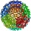 PDB-1z8y: 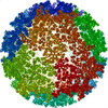 PDB-2xfb: 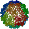 PDB-3muw: |
-原子モデル構築 2
| 初期モデル | PDB ID: |
|---|---|
| ソフトウェア | 名称: EMFIT |
| 詳細 | Protocol: Rigid body. each protein in the asymmetric unit was fitting individually. |
| 精密化 | 空間: REAL / プロトコル: RIGID BODY FIT / 当てはまり具合の基準: sumf |
| 得られたモデル | PDB-1z8y: PDB-2xfb: PDB-3muw: |