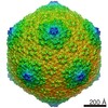
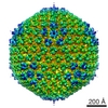
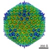
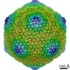
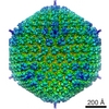
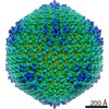
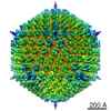
ジャーナル: EMBO J / 年: 2005 タイトル: A quasi-atomic model of human adenovirus type 5 capsid. 著者: Céline M S Fabry / Manuel Rosa-Calatrava / James F Conway / Chloé Zubieta / Stephen Cusack / Rob W H Ruigrok / Guy Schoehn / 要旨: Adenoviruses infect a wide range of vertebrates including humans. Their icosahedral capsids are composed of three major proteins: the trimeric hexon forms the facets and the penton, a noncovalent ...Adenoviruses infect a wide range of vertebrates including humans. Their icosahedral capsids are composed of three major proteins: the trimeric hexon forms the facets and the penton, a noncovalent complex of the pentameric penton base and trimeric fibre proteins, is located at the 12 capsid vertices. Several proteins (IIIa, VI, VIII and IX) stabilise the capsid. We have obtained a 10 A resolution map of the human adenovirus 5 by image analysis from cryo-electron micrographs (cryoEMs). This map, in combination with the X-ray structures of the penton base and hexon, was used to build a quasi-atomic model of the arrangement of the two major capsid components and to analyse the hexon-hexon and hexon-penton interactions. The secondary proteins, notably VIII, were located by comparing cryoEM maps of native and pIX deletion mutant virions. Minor proteins IX and IIIa are located on the outside of the capsid, whereas protein VIII is organised with a T=2 lattice on the inner face of the capsid. The capsid organisation is compared with the known X-ray structure of bacteriophage PRD1.
想定した対称性 - 点群: I (正20面体型対称) / アルゴリズム: OTHER / 解像度のタイプ: BY AUTHOR / 解像度: 15.5 Å / 解像度の算出法: FSC 0.33 CUT-OFF / ソフトウェア - 名称: PFT EM3DR / 詳細: Final maps were calculated from 1060 single images / 使用した粒子像数: 1060
 ムービー
ムービー コントローラー


 データを開く
 基本情報
基本情報 マップデータ
 試料
試料 機能・相同性情報
 unidentified adenovirus (ウイルス)
 データ登録者
 引用
引用
 構造の表示
構造の表示 UCSF Chimera
 ダウンロードとリンク
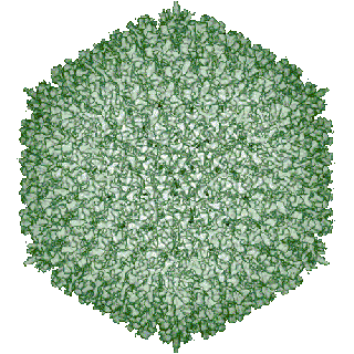 1112.gif
 http://ftp.pdbj.org/pub/emdb/structures/EMD-1112


 Y (Sec.)
 X (Row.)
 Z (Col.)


 試料の構成要素
 Homo sapiens (ヒト) / 別称: VERTEBRATES
 解析
解析 電子顕微鏡法