- EMDB-1063: Structure of the signal recognition particle interacting with the... -
+
Open data
ID or keywords:
Loading...
-
Basic information
Entry
Database: EMDB / ID: EMD-1063
Title
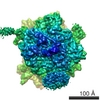
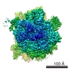
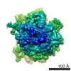
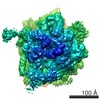
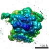
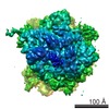
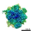
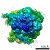
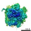
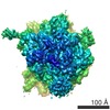
Structure of the signal recognition particle interacting with the elongation-arrested ribosome.
Map data
Structure of signal recognition particle interacting with elongation arested ribosome
Sample
Sample: 80S RNC-SRP complex fron canis sp.
Complex: 80S ribosome
Ligand: mammalian SRP
Ligand: tRNA
Function / homology
Function and homology information
signal recognition particle receptor complex / SRP-dependent cotranslational protein targeting to membrane, signal sequence recognition / endoplasmic reticulum signal sequence receptor activity / SRP-dependent cotranslational protein targeting to membrane / signal recognition particle, endoplasmic reticulum targeting / signal recognition particle / absorption of visible light / signal recognition particle binding / cotranslational protein targeting to membrane / G protein-coupled opsin signaling pathway ...signal recognition particle receptor complex / SRP-dependent cotranslational protein targeting to membrane, signal sequence recognition / endoplasmic reticulum signal sequence receptor activity / SRP-dependent cotranslational protein targeting to membrane / signal recognition particle, endoplasmic reticulum targeting / signal recognition particle / absorption of visible light / signal recognition particle binding / cotranslational protein targeting to membrane / G protein-coupled opsin signaling pathway / 11-cis retinal binding / G protein-coupled photoreceptor activity / photoreceptor inner segment membrane / granulocyte differentiation / signal-recognition-particle GTPase / negative regulation of translational elongation / protein targeting to ER / SRP-dependent cotranslational protein targeting to membrane, translocation / 7S RNA binding / SRP-dependent cotranslational protein targeting to membrane / exocrine pancreas development / photoreceptor outer segment membrane / SRP-dependent cotranslational protein targeting to membrane / ribonucleoprotein complex binding / neutrophil chemotaxis / visual perception / photoreceptor disc membrane / GDP binding / secretory granule lumen / ficolin-1-rich granule lumen / nuclear speck / nuclear body / GTPase activity / Neutrophil degranulation / GTP binding / nucleolus / endoplasmic reticulum / RNA binding / extracellular region / membrane / metal ion binding / nucleus / plasma membrane / cytoplasm / cytosol Similarity search - Function
Signal recognition particle, SRP9 subunit / SRP9 domain / Signal recognition particle SRP9 / Signal recognition particle 9 kDa protein (SRP9) / Signal recognition particle, SRP14 subunit / Signal recognition particle, SRP9/SRP14 subunit / Signal recognition particle 14kD protein / Signal recognition particle, SRP54 subunit, eukaryotic / Signal recognition particle, SRP19 subunit / Signal recognition particle, subunit SRP19-like superfamily ...Signal recognition particle, SRP9 subunit / SRP9 domain / Signal recognition particle SRP9 / Signal recognition particle 9 kDa protein (SRP9) / Signal recognition particle, SRP14 subunit / Signal recognition particle, SRP9/SRP14 subunit / Signal recognition particle 14kD protein / Signal recognition particle, SRP54 subunit, eukaryotic / Signal recognition particle, SRP19 subunit / Signal recognition particle, subunit SRP19-like superfamily / SRP19 protein / Signal recognition particle protein / Rhodopsin, N-terminal / Amino terminal of the G-protein receptor rhodopsin / Rhodopsin / SRP/SRP receptor, N-terminal / Signal recognition particle, SRP54 subunit / Signal recognition particle, SRP54 subunit, M-domain / Signal recognition particle, SRP54 subunit, M-domain superfamily / Signal peptide binding domain / Opsin / Visual pigments (opsins) retinal binding site / Visual pigments (opsins) retinal binding site. / SRP54-type proteins GTP-binding domain signature. / : / Signal recognition particle SRP54, helical bundle / Signal recognition particle SRP54, N-terminal domain superfamily / SRP54-type protein, helical bundle domain / SRP54-type protein, helical bundle domain / Signal recognition particle, SRP54 subunit, GTPase domain / SRP54-type protein, GTPase domain / SRP54-type protein, GTPase domain / Serpentine type 7TM GPCR chemoreceptor Srsx / G-protein coupled receptors family 1 signature. / 7 transmembrane receptor (rhodopsin family) / G protein-coupled receptor, rhodopsin-like / GPCR, rhodopsin-like, 7TM / G-protein coupled receptors family 1 profile. / ATPases associated with a variety of cellular activities / AAA+ ATPase domain / P-loop containing nucleoside triphosphate hydrolase Similarity search - Domain/homology
Signal recognition particle protein / Rhodopsin / Signal recognition particle 19 kDa protein / Signal recognition particle subunit SRP54 / Signal recognition particle 14 kDa protein / Signal recognition particle 9 kDa protein Similarity search - Component
Biological species
Canis lupus familiaris (dog)
Method
single particle reconstruction / cryo EM / Resolution: 12.0 Å
Journal: Nature / Year: 2004 Title: Structure of the signal recognition particle interacting with the elongation-arrested ribosome. Authors: Mario Halic / Thomas Becker / Martin R Pool / Christian M T Spahn / Robert A Grassucci / Joachim Frank / Roland Beckmann / Abstract: Cotranslational translocation of proteins across or into membranes is a vital process in all kingdoms of life. It requires that the translating ribosome be targeted to the membrane by the signal ...Cotranslational translocation of proteins across or into membranes is a vital process in all kingdoms of life. It requires that the translating ribosome be targeted to the membrane by the signal recognition particle (SRP), an evolutionarily conserved ribonucleoprotein particle. SRP recognizes signal sequences of nascent protein chains emerging from the ribosome. Subsequent binding of SRP leads to a pause in peptide elongation and to the ribosome docking to the membrane-bound SRP receptor. Here we present the structure of a targeting complex consisting of mammalian SRP bound to an active 80S ribosome carrying a signal sequence. This structure, solved to 12 A by cryo-electron microscopy, enables us to generate a molecular model of SRP in its functional conformation. The model shows how the S domain of SRP contacts the large ribosomal subunit at the nascent chain exit site to bind the signal sequence, and that the Alu domain reaches into the elongation-factor-binding site of the ribosome, explaining its elongation arrest activity.
History
Deposition
Dec 19, 2003
-
Header (metadata) release
Dec 19, 2003
-
Map release
Dec 19, 2004
-
Update
May 26, 2011
-
Current status
May 26, 2011
Processing site: PDBe / Status: Released
-
Structure visualization
Movie
Surface view with section colored by density value
In the structure databanks used in Yorodumi, some data are registered as the other names, "COVID-19 virus" and "2019-nCoV". Here are the details of the virus and the list of structure data.
Jan 31, 2019. EMDB accession codes are about to change! (news from PDBe EMDB page)
EMDB accession codes are about to change! (news from PDBe EMDB page)
The allocation of 4 digits for EMDB accession codes will soon come to an end. Whilst these codes will remain in use, new EMDB accession codes will include an additional digit and will expand incrementally as the available range of codes is exhausted. The current 4-digit format prefixed with “EMD-” (i.e. EMD-XXXX) will advance to a 5-digit format (i.e. EMD-XXXXX), and so on. It is currently estimated that the 4-digit codes will be depleted around Spring 2019, at which point the 5-digit format will come into force.
The EM Navigator/Yorodumi systems omit the EMD- prefix.
Related info.:Q: What is EMD? / ID/Accession-code notation in Yorodumi/EM Navigator
Yorodumi is a browser for structure data from EMDB, PDB, SASBDB, etc.
This page is also the successor to EM Navigator detail page, and also detail information page/front-end page for Omokage search.
The word "yorodu" (or yorozu) is an old Japanese word meaning "ten thousand". "mi" (miru) is to see.
Related info.:EMDB / PDB / SASBDB / Comparison of 3 databanks / Yorodumi Search / Aug 31, 2016. New EM Navigator & Yorodumi / Yorodumi Papers / Jmol/JSmol / Function and homology information / Changes in new EM Navigator and Yorodumi
 Movie
Movie Controller

 Yorodumi
 Open data
 Basic information
 Map data
 Sample
 Function and homology information
 Authors
 Citation
 Structure visualization
 Downloads & links
 1063.gif
 http://ftp.pdbj.org/pub/emdb/structures/EMD-1063


 Z (Sec.)
 Y (Row.)
 X (Col.)


 Sample components
 wheat germ (plant)
 Processing
 Electron microscopy
 FIELD EMISSION GUN
