+ Open data
- Basic information
| Entry | Database: EMDB / ID: EMD-6103 | |||||||||
|---|---|---|---|---|---|---|---|---|---|---|
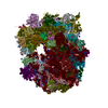
| Title | Cryo-EM Structure of the 80S-RAC complex from yeast | |||||||||
 Map data Map data | Reconstruction of yeast 80S-RAC complex, (All particles in Dataset RAC6) | |||||||||
 Sample Sample |
| |||||||||
 Keywords Keywords | ribosome-associated complex / co-translational chaperone / cryo-em / translation regulation / Zuotin / Ssz | |||||||||
| Biological species |  | |||||||||
| Method | single particle reconstruction / cryo EM / Resolution: 10.2 Å | |||||||||
 Authors Authors | Zhang YX / Ma CY / Yuan Y / Zhu J / Li NN / Chen C / Wu S / Yu L / Lei JL / Gao N | |||||||||
 Citation Citation |  Journal: Nat Struct Mol Biol / Year: 2014 Journal: Nat Struct Mol Biol / Year: 2014Title: Structural basis for interaction of a cotranslational chaperone with the eukaryotic ribosome. Authors: Yixiao Zhang / Chengying Ma / Yi Yuan / Jing Zhu / Ningning Li / Chu Chen / Shan Wu / Li Yu / Jianlin Lei / Ning Gao /  Abstract: Cotranslational chaperones, ubiquitous in all living organisms, protect nascent polypeptides from aggregation and facilitate their de novo folding. Importantly, emerging data have also suggested that ...Cotranslational chaperones, ubiquitous in all living organisms, protect nascent polypeptides from aggregation and facilitate their de novo folding. Importantly, emerging data have also suggested that ribosome-associated cotranslational chaperones have active regulatory roles in modulating protein translation. By characterizing the structure of a type of eukaryotic cotranslational chaperone, the ribosome-associated complex (RAC) from Saccharomyces cerevisiae, we show that RAC cross-links two ribosomal subunits, through a single long α-helix, to limit the predominant intersubunit rotation required for peptide elongation. We further demonstrate that any changes in the continuity, length or rigidity of this middle α-helix impair RAC function in vivo. Our results suggest a new mechanism in which RAC directly regulates protein translation by mechanically coupling cotranslational folding with the peptide-elongation cycle, and they lay the foundation for further exploration of regulatory roles of RAC in translation control. | |||||||||
| History |
|
- Structure visualization
| Movie |
 Movie viewer Movie viewer |
|---|---|
| Structure viewer | EM map:  SurfView SurfView Molmil Molmil Jmol/JSmol Jmol/JSmol |
| Supplemental images |
- Downloads & links
-EMDB archive
| Map data |  emd_6103.map.gz emd_6103.map.gz | 16.8 MB |  EMDB map data format EMDB map data format | |
|---|---|---|---|---|
| Header (meta data) |  emd-6103-v30.xml emd-6103-v30.xml emd-6103.xml emd-6103.xml | 9.6 KB 9.6 KB | Display Display |  EMDB header EMDB header |
| FSC (resolution estimation) |  emd_6103_fsc.xml emd_6103_fsc.xml | 7 KB | Display |  FSC data file FSC data file |
| Images |  emd_6103.png emd_6103.png | 831 KB | ||
| Archive directory |  http://ftp.pdbj.org/pub/emdb/structures/EMD-6103 http://ftp.pdbj.org/pub/emdb/structures/EMD-6103 ftp://ftp.pdbj.org/pub/emdb/structures/EMD-6103 ftp://ftp.pdbj.org/pub/emdb/structures/EMD-6103 | HTTPS FTP |
-Related structure data
- Links
Links
| EMDB pages |  EMDB (EBI/PDBe) / EMDB (EBI/PDBe) /  EMDataResource EMDataResource |
|---|---|
| Related items in Molecule of the Month |
- Map
Map
| File |  Download / File: emd_6103.map.gz / Format: CCP4 / Size: 17.7 MB / Type: IMAGE STORED AS FLOATING POINT NUMBER (4 BYTES) Download / File: emd_6103.map.gz / Format: CCP4 / Size: 17.7 MB / Type: IMAGE STORED AS FLOATING POINT NUMBER (4 BYTES) | ||||||||||||||||||||||||||||||||||||||||||||||||||||||||||||
|---|---|---|---|---|---|---|---|---|---|---|---|---|---|---|---|---|---|---|---|---|---|---|---|---|---|---|---|---|---|---|---|---|---|---|---|---|---|---|---|---|---|---|---|---|---|---|---|---|---|---|---|---|---|---|---|---|---|---|---|---|---|
| Annotation | Reconstruction of yeast 80S-RAC complex, (All particles in Dataset RAC6) | ||||||||||||||||||||||||||||||||||||||||||||||||||||||||||||
| Projections & slices | Image control
Images are generated by Spider. | ||||||||||||||||||||||||||||||||||||||||||||||||||||||||||||
| Voxel size | X=Y=Z: 2.68 Å | ||||||||||||||||||||||||||||||||||||||||||||||||||||||||||||
| Density |
| ||||||||||||||||||||||||||||||||||||||||||||||||||||||||||||
| Symmetry | Space group: 1 | ||||||||||||||||||||||||||||||||||||||||||||||||||||||||||||
| Details | EMDB XML:
CCP4 map header:
| ||||||||||||||||||||||||||||||||||||||||||||||||||||||||||||
-Supplemental data
- Sample components
-Entire : Saccharomyces cerevisiae 80S ribosome bound with RAC
| Entire | Name: Saccharomyces cerevisiae 80S ribosome bound with RAC |
|---|---|
| Components |
|
-Supramolecule #1000: Saccharomyces cerevisiae 80S ribosome bound with RAC
| Supramolecule | Name: Saccharomyces cerevisiae 80S ribosome bound with RAC / type: sample / ID: 1000 / Number unique components: 2 |
|---|
-Supramolecule #1: Saccharomyces cerevisiae 80S ribosome
| Supramolecule | Name: Saccharomyces cerevisiae 80S ribosome / type: complex / ID: 1 / Recombinant expression: Yes / Ribosome-details: ribosome-eukaryote: ALL |
|---|---|
| Source (natural) | Organism:  |
| Recombinant expression | Organism:  |
-Macromolecule #1: Ribosome-associated complex (RAC)
| Macromolecule | Name: Ribosome-associated complex (RAC) / type: protein_or_peptide / ID: 1 / Recombinant expression: Yes |
|---|---|
| Source (natural) | Organism:  |
| Recombinant expression | Organism:  |
-Experimental details
-Structure determination
| Method | cryo EM |
|---|---|
 Processing Processing | single particle reconstruction |
| Aggregation state | particle |
- Sample preparation
| Buffer | pH: 7.4 / Details: 20 mM Hepes-KOH, 120 mM KOAc, 10 mM Mg(OAc)2 |
|---|---|
| Vitrification | Cryogen name: ETHANE / Chamber humidity: 90 % / Instrument: FEI VITROBOT MARK IV |
- Electron microscopy
| Microscope | FEI TECNAI F20 |
|---|---|
| Date | Aug 3, 2012 |
| Image recording | Category: CCD / Film or detector model: GATAN ULTRASCAN 4000 (4k x 4k) / Number real images: 1721 / Average electron dose: 20 e/Å2 |
| Electron beam | Acceleration voltage: 200 kV / Electron source:  FIELD EMISSION GUN FIELD EMISSION GUN |
| Electron optics | Illumination mode: FLOOD BEAM / Imaging mode: BRIGHT FIELD |
| Sample stage | Specimen holder model: GATAN LIQUID NITROGEN |
| Experimental equipment |  Model: Tecnai F20 / Image courtesy: FEI Company |
 Movie
Movie Controller


 Z (Sec.)
 Y (Row.)
 X (Col.)


