 Movie
Movie Controller


+ Open data

- Basic information
| Entry | Database: EMDB / ID: EMD-1768 | |||||||||
|---|---|---|---|---|---|---|---|---|---|---|
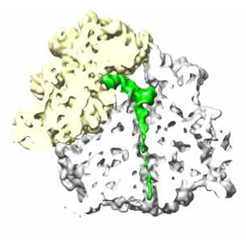
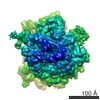
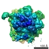
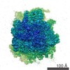
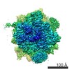
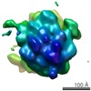
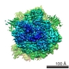
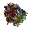
| Title | CMV-stalled wheat germ 80S ribosome | |||||||||
 Map data Map data | This map represents a CMV (cytomegalovirus) stalled wheat germ 80S ribosome | |||||||||
 Sample Sample |
| |||||||||
 Keywords Keywords | Antibiotic / ribosome / translation / stalling / cytomegalovirus | |||||||||
| Biological species |  | |||||||||
| Method | single particle reconstruction / cryo EM / negative staining / Resolution: 6.5 Å | |||||||||
 Authors Authors | Bhushan S / Meyer H / Starosta A / Becker T / Mielke T / Berninghausen O / Sattler M / Wilson D / Beckmann R | |||||||||
 Citation Citation | Journal: Mol Cell / Year: 2010 Title: Structural basis for translational stalling by human cytomegalovirus and fungal arginine attenuator peptide. Authors: Shashi Bhushan / Helge Meyer / Agata L Starosta / Thomas Becker / Thorsten Mielke / Otto Berninghausen / Michael Sattler / Daniel N Wilson / Roland Beckmann /  Abstract: Specific regulatory nascent chains establish direct interactions with the ribosomal tunnel, leading to translational stalling. Despite a wealth of biochemical data, structural insight into the ...Specific regulatory nascent chains establish direct interactions with the ribosomal tunnel, leading to translational stalling. Despite a wealth of biochemical data, structural insight into the mechanism of translational stalling in eukaryotes is still lacking. Here we use cryo-electron microscopy to visualize eukaryotic ribosomes stalled during the translation of two diverse regulatory peptides: the fungal arginine attenuator peptide (AAP) and the human cytomegalovirus (hCMV) gp48 upstream open reading frame 2 (uORF2). The C terminus of the AAP appears to be compacted adjacent to the peptidyl transferase center (PTC). Both nascent chains interact with ribosomal proteins L4 and L17 at tunnel constriction in a distinct fashion. Significant changes at the PTC were observed: the eukaryotic-specific loop of ribosomal protein L10e establishes direct contact with the CCA end of the peptidyl-tRNA (P-tRNA), which may be critical for silencing of the PTC during translational stalling. Our findings provide direct structural insight into two distinct eukaryotic stalling processes. | |||||||||
| History |
|
- Structure visualization
| Movie |
 Movie viewer Movie viewer |
|---|---|
| Structure viewer | EM map: SurfViewMolmilJmol/JSmol |
| Supplemental images |
- Downloads & links
-EMDB archive
| Map data | emd_1768.map.gz | 30.4 MB | EMDB map data format | |
|---|---|---|---|---|
| Header (meta data) | emd-1768-v30.xmlemd-1768.xml | 9.5 KB 9.5 KB | Display Display | EMDB header |
| Images |  CMV_RNC.jpg CMV_RNC.jpg | 129.6 KB | ||
| Archive directory |  http://ftp.pdbj.org/pub/emdb/structures/EMD-1768ftp://ftp.pdbj.org/pub/emdb/structures/EMD-1768 http://ftp.pdbj.org/pub/emdb/structures/EMD-1768ftp://ftp.pdbj.org/pub/emdb/structures/EMD-1768 | HTTPS FTP |
-Related structure data

-Links
| EMDB pages | EMDB (EBI/PDBe) / EMDataResource |
|---|---|
| Related items in Molecule of the Month |

-Map
| File | Download / File: emd_1768.map.gz / Format: CCP4 / Size: 185.7 MB / Type: IMAGE STORED AS FLOATING POINT NUMBER (4 BYTES) | ||||||||||||||||||||||||||||||||||||||||||||||||||||||||||||||||||||
|---|---|---|---|---|---|---|---|---|---|---|---|---|---|---|---|---|---|---|---|---|---|---|---|---|---|---|---|---|---|---|---|---|---|---|---|---|---|---|---|---|---|---|---|---|---|---|---|---|---|---|---|---|---|---|---|---|---|---|---|---|---|---|---|---|---|---|---|---|---|
| Annotation | This map represents a CMV (cytomegalovirus) stalled wheat germ 80S ribosome | ||||||||||||||||||||||||||||||||||||||||||||||||||||||||||||||||||||
| Projections & slices | Image control
Images are generated by Spider. | ||||||||||||||||||||||||||||||||||||||||||||||||||||||||||||||||||||
| Voxel size | X=Y=Z: 1.2375 Å | ||||||||||||||||||||||||||||||||||||||||||||||||||||||||||||||||||||
| Density |
| ||||||||||||||||||||||||||||||||||||||||||||||||||||||||||||||||||||
| Symmetry | Space group: 1 | ||||||||||||||||||||||||||||||||||||||||||||||||||||||||||||||||||||
| Details | EMDB XML:
CCP4 map header:
| ||||||||||||||||||||||||||||||||||||||||||||||||||||||||||||||||||||
 Z (Sec.)
 X (Row.)
 Y (Col.)


-Supplemental data
- Sample components
-Entire : hCMV-stalled wheat germ 80S ribosome
| Entire | Name: hCMV-stalled wheat germ 80S ribosome |
|---|---|
| Components |
|
-Supramolecule #1000: hCMV-stalled wheat germ 80S ribosome
| Supramolecule | Name: hCMV-stalled wheat germ 80S ribosome / type: sample / ID: 1000 / Details: Single particle / Oligomeric state: One ribosome / Number unique components: 1 |
|---|---|
| Molecular weight | Experimental: 4.2 MDa / Theoretical: 4.2 MDa |
-Supramolecule #1: T. aestivum 80S ribosome
| Supramolecule | Name: T. aestivum 80S ribosome / type: complex / ID: 1 / Name.synonym: Wheat germ ribosome / Recombinant expression: No / Ribosome-details: ribosome-eukaryote: ALL |
|---|---|
| Source (natural) | Organism: |
| Molecular weight | Experimental: 4.2 MDa / Theoretical: 4.2 MDa |
-Experimental details
-Structure determination
| Method | negative staining, cryo EM |
|---|---|
 Processing Processing | single particle reconstruction |
| Aggregation state | particle |
-Sample preparation
| Concentration | 0.02 mg/mL |
|---|---|
| Buffer | pH: 7.5 Details: 30 mM HEPES/KOH, pH 7.5 180 mM KOAc, 10 mM Mg(OAc)2, 0.01 mg/ml cycloheximide, 1 mM DTT,3.5 % (w/v) glycerol 0.3 % (w/v) digitonin |
| Staining | Type: NEGATIVE / Details: Cryo-EM |
| Grid | Details: Quantifoil Grid with 2 nm carbon on top |
| Vitrification | Cryogen name: ETHANE / Chamber humidity: 100 % / Instrument: OTHER / Details: Vitrification instrument: Vitrobot Method: Blot for 10 seconds before plunging, use 2 layers of filter paper |
- Electron microscopy
| Microscope | FEI POLARA 300 |
|---|---|
| Specialist optics | Energy filter - Name: FEI |
| Image recording | Category: FILM / Film or detector model: KODAK SO-163 FILM / Digitization - Scanner: OTHER / Digitization - Sampling interval: 4.76 µm / Average electron dose: 25 e/Å2 Details: Scanned at 5334 dpi on a Heidelberg Primescan Drum Scanner Od range: 1.2 |
| Electron beam | Acceleration voltage: 300 kV / Electron source:  FIELD EMISSION GUN FIELD EMISSION GUN |
| Electron optics | Calibrated magnification: 38000 / Illumination mode: FLOOD BEAM / Imaging mode: BRIGHT FIELD / Cs: 2.26 mm / Nominal defocus max: 4.5 µm / Nominal defocus min: 1.0 µm / Nominal magnification: 39000 |
| Sample stage | Specimen holder: FEI Polara Cartridge System / Specimen holder model: OTHER |
| Experimental equipment |  Model: Tecnai Polara / Image courtesy: FEI Company |
-Image processing
| Details | The nascent polypeptide chain was saturated with mammalian Sec61 (see Becker et al., Science 2009) to avoid orientational bias on the grid. |
|---|---|
| CTF correction | Details: SPIDER TF CTS |
| Final reconstruction | Applied symmetry - Point group: C1 (asymmetric) / Algorithm: OTHER / Resolution.type: BY AUTHOR / Resolution: 6.5 Å / Resolution method: FSC 0.5 CUT-OFF / Software - Name: SPIDER / Number images used: 150000 |
