- EMDB-1063: Structure of the signal recognition particle interacting with the... -
+
データを開く
IDまたはキーワード:
読み込み中...
-
基本情報
登録情報
データベース: EMDB / ID: EMD-1063
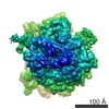
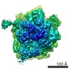
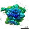
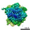
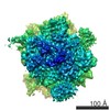
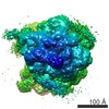
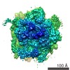
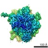
タイトル
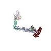
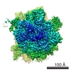
Structure of the signal recognition particle interacting with the elongation-arrested ribosome.
マップデータ
Structure of signal recognition particle interacting with elongation arested ribosome
試料
試料: 80S RNC-SRP complex fron canis sp.
複合体: 80S ribosomeEukaryotic ribosome
リガンド: mammalian SRP
リガンド: tRNA転移RNA
機能・相同性
機能・相同性情報
signal recognition particle receptor complex / SRP-dependent cotranslational protein targeting to membrane / SRP-dependent cotranslational protein targeting to membrane, signal sequence recognition / endoplasmic reticulum signal peptide binding / signal recognition particle, endoplasmic reticulum targeting / signal recognition particle binding / granulocyte differentiation / absorption of visible light / negative regulation of translational elongation / G protein-coupled photoreceptor activity ...signal recognition particle receptor complex / SRP-dependent cotranslational protein targeting to membrane / SRP-dependent cotranslational protein targeting to membrane, signal sequence recognition / endoplasmic reticulum signal peptide binding / signal recognition particle, endoplasmic reticulum targeting / signal recognition particle binding / granulocyte differentiation / absorption of visible light / negative regulation of translational elongation / G protein-coupled photoreceptor activity / シグナル認識粒子 / photoreceptor inner segment membrane / rhodopsin mediated signaling pathway / 11-cis retinal binding / cotranslational protein targeting to membrane / signal-recognition-particle GTPase / protein targeting to ER / SRP-dependent cotranslational protein targeting to membrane, translocation / 7S RNA binding / exocrine pancreas development / SRP-dependent cotranslational protein targeting to membrane / photoreceptor outer segment membrane / SRP-dependent cotranslational protein targeting to membrane / ribonucleoprotein complex binding / 視覚 / 好中球 / photoreceptor disc membrane / GDP binding / secretory granule lumen / ficolin-1-rich granule lumen / nuclear body / nuclear speck / GTPase activity / Neutrophil degranulation / GTP binding / 核小体 / 小胞体 / ATP hydrolysis activity / RNA binding / extracellular region / 生体膜 / metal ion binding / 細胞核 / 細胞膜 / 細胞質基質 / 細胞質 類似検索 - 分子機能
Signal recognition particle, SRP9 subunit / SRP9 domain / Signal recognition particle SRP9 / Signal recognition particle 9 kDa protein (SRP9) / Signal recognition particle, SRP14 subunit / Signal recognition particle, SRP9/SRP14 subunit / Signal recognition particle 14kD protein / Signal recognition particle, SRP54 subunit, eukaryotic / Signal recognition particle, SRP19 subunit / Signal recognition particle, subunit SRP19-like superfamily ...Signal recognition particle, SRP9 subunit / SRP9 domain / Signal recognition particle SRP9 / Signal recognition particle 9 kDa protein (SRP9) / Signal recognition particle, SRP14 subunit / Signal recognition particle, SRP9/SRP14 subunit / Signal recognition particle 14kD protein / Signal recognition particle, SRP54 subunit, eukaryotic / Signal recognition particle, SRP19 subunit / Signal recognition particle, subunit SRP19-like superfamily / SRP19 protein / Signal recognition particle protein / Rhodopsin, N-terminal / Amino terminal of the G-protein receptor rhodopsin / ロドプシン / Opsin / Visual pigments (opsins) retinal binding site / Visual pigments (opsins) retinal binding site. / SRP/SRP receptor, N-terminal / Signal recognition particle, SRP54 subunit / Signal recognition particle, SRP54 subunit, M-domain / Signal recognition particle, SRP54 subunit, M-domain superfamily / Signal peptide binding domain / SRP54-type proteins GTP-binding domain signature. / Signal recognition particle SRP54, helical bundle / Signal recognition particle SRP54, N-terminal domain superfamily / SRP54-type protein, helical bundle domain / SRP54-type protein, helical bundle domain / Signal recognition particle, SRP54 subunit, GTPase domain / SRP54-type protein, GTPase domain / SRP54-type protein, GTPase domain / Serpentine type 7TM GPCR chemoreceptor Srsx / G-protein coupled receptors family 1 signature. / G protein-coupled receptor, rhodopsin-like / GPCR, rhodopsin-like, 7TM / G-protein coupled receptors family 1 profile. / 7 transmembrane receptor (rhodopsin family) / ATPases associated with a variety of cellular activities / AAA+ ATPase domain / P-loop containing nucleoside triphosphate hydrolase 類似検索 - ドメイン・相同性
Signal recognition particle protein / ロドプシン / Signal recognition particle 19 kDa protein / Signal recognition particle subunit SRP54 / Signal recognition particle 14 kDa protein / Signal recognition particle 9 kDa protein 類似検索 - 構成要素
ジャーナル: Nature / 年: 2004 タイトル: Structure of the signal recognition particle interacting with the elongation-arrested ribosome. 著者: Mario Halic / Thomas Becker / Martin R Pool / Christian M T Spahn / Robert A Grassucci / Joachim Frank / Roland Beckmann / 要旨: Cotranslational translocation of proteins across or into membranes is a vital process in all kingdoms of life. It requires that the translating ribosome be targeted to the membrane by the signal ...Cotranslational translocation of proteins across or into membranes is a vital process in all kingdoms of life. It requires that the translating ribosome be targeted to the membrane by the signal recognition particle (SRP), an evolutionarily conserved ribonucleoprotein particle. SRP recognizes signal sequences of nascent protein chains emerging from the ribosome. Subsequent binding of SRP leads to a pause in peptide elongation and to the ribosome docking to the membrane-bound SRP receptor. Here we present the structure of a targeting complex consisting of mammalian SRP bound to an active 80S ribosome carrying a signal sequence. This structure, solved to 12 A by cryo-electron microscopy, enables us to generate a molecular model of SRP in its functional conformation. The model shows how the S domain of SRP contacts the large ribosomal subunit at the nascent chain exit site to bind the signal sequence, and that the Alu domain reaches into the elongation-factor-binding site of the ribosome, explaining its elongation arrest activity.
 ムービー
ムービー コントローラー


 データを開く
 基本情報
基本情報 マップデータ
 試料
試料 機能・相同性情報
 signal recognition particle receptor complex / SRP-dependent cotranslational protein targeting to membrane / SRP-dependent cotranslational protein targeting to membrane, signal sequence recognition / endoplasmic reticulum signal peptide binding /
 データ登録者
 引用
引用
 構造の表示
構造の表示
 ダウンロードとリンク
 1063.gif
 http://ftp.pdbj.org/pub/emdb/structures/EMD-1063
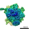


 試料の構成要素
 解析
解析 電子顕微鏡法