+ データを開く
- 基本情報
基本情報
| 登録情報 | データベース: EMDB / ID: EMD-0233 | |||||||||
|---|---|---|---|---|---|---|---|---|---|---|
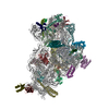
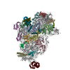
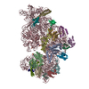
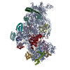
| タイトル | Cryo-EM structure of the Trypanosoma brucei mitochondrial ribosome - This entry contains the head of the small mitoribosomal subunit | |||||||||
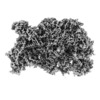
 マップデータ マップデータ | map of the head of the T. brucei mitoribosome small subunit | |||||||||
 試料 試料 |
| |||||||||
 キーワード キーワード | mitoribosome / translation / Trypanosoma / small ribosomal subunit / 9S rRNA / ribosomal protein / RIBOSOME | |||||||||
| 機能・相同性 |  機能・相同性情報 機能・相同性情報mitochondrial mRNA editing complex / mitochondrial RNA processing / kinetoplast / thiosulfate sulfurtransferase activity / nuclear lumen / ciliary plasm / mRNA stabilization / mitochondrial small ribosomal subunit / RNA processing / mitochondrion organization ...mitochondrial mRNA editing complex / mitochondrial RNA processing / kinetoplast / thiosulfate sulfurtransferase activity / nuclear lumen / ciliary plasm / mRNA stabilization / mitochondrial small ribosomal subunit / RNA processing / mitochondrion organization / structural constituent of ribosome / translation / mRNA binding / mitochondrion / RNA binding / nucleoplasm / nucleus / cytoplasm 類似検索 - 分子機能 | |||||||||
| 生物種 |  | |||||||||
| 手法 | 単粒子再構成法 / クライオ電子顕微鏡法 / 解像度: 3.08 Å | |||||||||
 データ登録者 データ登録者 | Ramrath DJF / Niemann M | |||||||||
| 資金援助 |  スイス, 1件 スイス, 1件
| |||||||||
 引用 引用 |  ジャーナル: Science / 年: 2018 ジャーナル: Science / 年: 2018タイトル: Evolutionary shift toward protein-based architecture in trypanosomal mitochondrial ribosomes. 著者: David J F Ramrath / Moritz Niemann / Marc Leibundgut / Philipp Bieri / Céline Prange / Elke K Horn / Alexander Leitner / Daniel Boehringer / André Schneider / Nenad Ban /  要旨: Ribosomal RNA (rRNA) plays key functional and architectural roles in ribosomes. Using electron microscopy, we determined the atomic structure of a highly divergent ribosome found in mitochondria of , ...Ribosomal RNA (rRNA) plays key functional and architectural roles in ribosomes. Using electron microscopy, we determined the atomic structure of a highly divergent ribosome found in mitochondria of , a unicellular parasite that causes sleeping sickness in humans. The trypanosomal mitoribosome features the smallest rRNAs and contains more proteins than all known ribosomes. The structure shows how the proteins have taken over the role of architectural scaffold from the rRNA: They form an autonomous outer shell that surrounds the entire particle and stabilizes and positions the functionally important regions of the rRNA. Our results also reveal the "minimal" set of conserved rRNA and protein components shared by all ribosomes that help us define the most essential functional elements. | |||||||||
| 履歴 |
|
- 構造の表示
構造の表示
| ムービー |
 ムービービューア ムービービューア |
|---|---|
| 構造ビューア | EMマップ:  SurfView SurfView Molmil Molmil Jmol/JSmol Jmol/JSmol |
| 添付画像 |
- ダウンロードとリンク
-EMDBアーカイブ
| マップデータ |  emd_0233.map.gz emd_0233.map.gz | 12.5 MB |  EMDBマップデータ形式 EMDBマップデータ形式 | |
|---|---|---|---|---|
| ヘッダ (付随情報) |  emd-0233-v30.xml emd-0233-v30.xml emd-0233.xml emd-0233.xml | 51.3 KB 51.3 KB | 表示 表示 |  EMDBヘッダ EMDBヘッダ |
| 画像 |  emd_0233.png emd_0233.png | 66.2 KB | ||
| Filedesc metadata |  emd-0233.cif.gz emd-0233.cif.gz | 15.4 KB | ||
| その他 |  emd_0233_half_map_1.map.gz emd_0233_half_map_1.map.gz emd_0233_half_map_2.map.gz emd_0233_half_map_2.map.gz | 97.9 MB 97.9 MB | ||
| アーカイブディレクトリ |  http://ftp.pdbj.org/pub/emdb/structures/EMD-0233 http://ftp.pdbj.org/pub/emdb/structures/EMD-0233 ftp://ftp.pdbj.org/pub/emdb/structures/EMD-0233 ftp://ftp.pdbj.org/pub/emdb/structures/EMD-0233 | HTTPS FTP |
-検証レポート
| 文書・要旨 |  emd_0233_validation.pdf.gz emd_0233_validation.pdf.gz | 365.3 KB | 表示 |  EMDB検証レポート EMDB検証レポート |
|---|---|---|---|---|
| 文書・詳細版 |  emd_0233_full_validation.pdf.gz emd_0233_full_validation.pdf.gz | 364.4 KB | 表示 | |
| XML形式データ |  emd_0233_validation.xml.gz emd_0233_validation.xml.gz | 12.4 KB | 表示 | |
| アーカイブディレクトリ |  https://ftp.pdbj.org/pub/emdb/validation_reports/EMD-0233 https://ftp.pdbj.org/pub/emdb/validation_reports/EMD-0233 ftp://ftp.pdbj.org/pub/emdb/validation_reports/EMD-0233 ftp://ftp.pdbj.org/pub/emdb/validation_reports/EMD-0233 | HTTPS FTP |
-関連構造データ
- リンク
リンク
| EMDBのページ |  EMDB (EBI/PDBe) / EMDB (EBI/PDBe) /  EMDataResource EMDataResource |
|---|---|
| 「今月の分子」の関連する項目 |
- マップ
マップ
| ファイル |  ダウンロード / ファイル: emd_0233.map.gz / 形式: CCP4 / 大きさ: 125 MB / タイプ: IMAGE STORED AS FLOATING POINT NUMBER (4 BYTES) ダウンロード / ファイル: emd_0233.map.gz / 形式: CCP4 / 大きさ: 125 MB / タイプ: IMAGE STORED AS FLOATING POINT NUMBER (4 BYTES) | ||||||||||||||||||||||||||||||||||||||||||||||||||||||||||||
|---|---|---|---|---|---|---|---|---|---|---|---|---|---|---|---|---|---|---|---|---|---|---|---|---|---|---|---|---|---|---|---|---|---|---|---|---|---|---|---|---|---|---|---|---|---|---|---|---|---|---|---|---|---|---|---|---|---|---|---|---|---|
| 注釈 | map of the head of the T. brucei mitoribosome small subunit | ||||||||||||||||||||||||||||||||||||||||||||||||||||||||||||
| 投影像・断面図 | 画像のコントロール
画像は Spider により作成 | ||||||||||||||||||||||||||||||||||||||||||||||||||||||||||||
| ボクセルのサイズ | X=Y=Z: 1.39 Å | ||||||||||||||||||||||||||||||||||||||||||||||||||||||||||||
| 密度 |
| ||||||||||||||||||||||||||||||||||||||||||||||||||||||||||||
| 対称性 | 空間群: 1 | ||||||||||||||||||||||||||||||||||||||||||||||||||||||||||||
| 詳細 | EMDB XML:
CCP4マップ ヘッダ情報:
| ||||||||||||||||||||||||||||||||||||||||||||||||||||||||||||
-添付データ
-ハーフマップ: half map (even) of the head of the...
| ファイル | emd_0233_half_map_1.map | ||||||||||||
|---|---|---|---|---|---|---|---|---|---|---|---|---|---|
| 注釈 | half map (even) of the head of the T. brucei mitoribosome small subunit | ||||||||||||
| 投影像・断面図 |
| ||||||||||||
| 密度ヒストグラム |
-ハーフマップ: half map (odd) of the head of the...
| ファイル | emd_0233_half_map_2.map | ||||||||||||
|---|---|---|---|---|---|---|---|---|---|---|---|---|---|
| 注釈 | half map (odd) of the head of the T. brucei mitoribosome small subunit | ||||||||||||
| 投影像・断面図 |
| ||||||||||||
| 密度ヒストグラム |
- 試料の構成要素
+全体 : head of the T. brucei mitoribosome small subunit
+超分子 #1: head of the T. brucei mitoribosome small subunit
+分子 #1: mS48
+分子 #2: mS59
+分子 #3: mS49
+分子 #4: mS50
+分子 #5: mS52
+分子 #6: mS53
+分子 #7: mS54
+分子 #8: mS55
+分子 #9: mS57
+分子 #10: mS58
+分子 #11: mS67
+分子 #12: mS69
+分子 #13: mS70
+分子 #14: mS71
+分子 #15: mS72
+分子 #16: uS3m
+分子 #17: uS9m
+分子 #18: uS10m
+分子 #19: uS11m
+分子 #20: uS14m
+分子 #21: uS18m
+分子 #22: uS19m
+分子 #23: mS29
+分子 #24: mS33
+分子 #25: mS35
+分子 #27: Unknown protein
+分子 #28: Unknown protein
+分子 #26: RNA (143-MER)
+分子 #29: SPERMIDINE
+分子 #30: URIDINE 5'-TRIPHOSPHATE
+分子 #31: GUANOSINE-5'-TRIPHOSPHATE
+分子 #32: MAGNESIUM ION
+分子 #33: water
-実験情報
-構造解析
| 手法 | クライオ電子顕微鏡法 |
|---|---|
 解析 解析 | 単粒子再構成法 |
| 試料の集合状態 | particle |
- 試料調製
試料調製
| 緩衝液 | pH: 7.4 |
|---|---|
| 凍結 | 凍結剤: ETHANE-PROPANE / チャンバー内湿度: 98 % / チャンバー内温度: 278 K / 装置: FEI VITROBOT MARK IV |
- 電子顕微鏡法
| 顕微鏡 | FEI TITAN KRIOS |
|---|---|
| 撮影 | フィルム・検出器のモデル: FEI FALCON III (4k x 4k) 検出モード: INTEGRATING / 平均電子線量: 40.0 e/Å2 |
| 電子線 | 加速電圧: 300 kV / 電子線源:  FIELD EMISSION GUN FIELD EMISSION GUN |
| 電子光学系 | 照射モード: FLOOD BEAM / 撮影モード: BRIGHT FIELD |
| 実験機器 |  モデル: Titan Krios / 画像提供: FEI Company |
- 画像解析
画像解析
| 初期モデル | モデルのタイプ: OTHER / 詳細: initial model from 2D class averages in EMAN |
|---|---|
| 最終 再構成 | アルゴリズム: FOURIER SPACE / 解像度のタイプ: BY AUTHOR / 解像度: 3.08 Å / 解像度の算出法: FSC 0.143 CUT-OFF / ソフトウェア - 名称: RELION / 使用した粒子像数: 101308 |
| 初期 角度割当 | タイプ: OTHER |
| 最終 角度割当 | タイプ: MAXIMUM LIKELIHOOD |
-原子モデル構築 1
| 詳細 | We used Coot and O for initial model building and refined the structure using Phenix |
|---|---|
| 精密化 | プロトコル: AB INITIO MODEL / 温度因子: 45 |
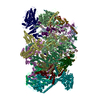
| 得られたモデル |  PDB-6hiz: |
 ムービー
ムービー コントローラー










 Z (Sec.)
 Y (Row.)
 X (Col.)


