[日本語] English
文献
- EMDB/PDB/SASBDBから引用されている文献のデータベース -

+
検索条件

キーワード
構造解析手法
著者・登録者
ジャーナル
IF

-
Structure paper

タイトルEvolutionary study and structural basis of proton sensing by Mus GPR4 and Xenopus GPR4.
ジャーナル・号・ページCell, Vol. 188, Issue 3, Page 653-670.e24, Year 2025
掲載日2025年2月6日
著者Xin Wen / Pan Shang / Haidi Chen / Lulu Guo / Naikang Rong / Xiaoyu Jiang / Xuan Li / Junyan Liu / Gongming Yang / Jiacheng Zhang / Kongkai Zhu / Qingbiao Meng / Xuefei He / Zhihai Wang / Zili Liu / Haoran Cheng / Yilin Zheng / Bifei Zhang / Jiaojiao Pang / Zhaoqian Liu / Peng Xiao / Yuguo Chen / Lunxu Liu / Fengming Luo / Xiao Yu / Fan Yi / Pengju Zhang / Fan Yang / Cheng Deng / Jin-Peng Sun /
PubMed 要旨Animals have evolved pH-sensing membrane receptors, such as G-protein-coupled receptor 4 (GPR4), to monitor pH changes related to their physiology and generate adaptive reactions. However, the ...Animals have evolved pH-sensing membrane receptors, such as G-protein-coupled receptor 4 (GPR4), to monitor pH changes related to their physiology and generate adaptive reactions. However, the evolutionary trajectory and structural mechanism of proton sensing by GPR4 remain unresolved. Here, we observed a positive correlation between the optimal pH of GPR4 activity and the blood pH range across different species. By solving 7-cryoelectron microscopy (cryo-EM) structures of Xenopus tropicalis GPR4 (xtGPR4) and Mus musculus GPR4 (mmGPR4) under varying pH conditions, we identified that protonation of H and H enabled polar network establishment and tighter association between the extracellular loop 2 (ECL2) and 7 transmembrane (7TM) domain, as well as a conserved propagating path, which are common mechanisms underlying protonation-induced GPR4 activation across different species. Moreover, protonation of distinct extracellular H contributed to the more acidic optimal pH range of xtGPR4. Overall, our study revealed common and distinct mechanisms of proton sensing by GPR4, from a structural, functional, and evolutionary perspective.
リンクCell / PubMed:39753131
手法EM (単粒子)
解像度2.36 - 3.78 Å
構造データ

EMDB-39946, PDB-8zd1:
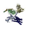
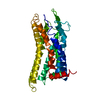
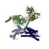
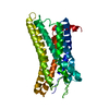
Cryo-EM structure of the xGPR4-Gs complex in pH6.2
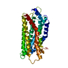
PDB-8zf4: Cryo-EM structure of the receptor of xGPR4-Gs complex in pH6.2
手法: EM (単粒子) / 解像度: 2.6 Å

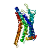
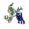
EMDB-60050, PDB-8zf4:
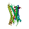
Cryo-EM structure of the receptor of xGPR4-Gs complex in pH6.2
PDB-8zd1: Cryo-EM structure of the xGPR4-Gs complex in pH6.2
手法: EM (単粒子) / 解像度: 3.05 Å

EMDB-60051, PDB-8zf6:
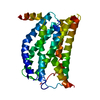
Cryo-EM structure of the xGPR4-Gs complex in pH6.7
PDB-8zf7: Cryo-EM structure of the receptor of xGPR4-Gs complex in pH6.7
手法: EM (単粒子) / 解像度: 2.98 Å

EMDB-60052, PDB-8zf7:
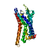
Cryo-EM structure of the receptor of xGPR4-Gs complex in pH6.7
手法: EM (単粒子) / 解像度: 3.15 Å

EMDB-60053, PDB-8zf9:
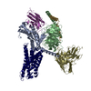
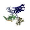
Cryo-EM structure of the mmGPR4-Gs complex in pH7.2
PDB-8zfe: Cryo-EM structure of the mmGPR4-Gs receptor in pH7.2
手法: EM (単粒子) / 解像度: 2.56 Å

EMDB-60054, PDB-8zfa:
Cryo-EM structure of the xtGPR4-Gs complex in pH7.2
PDB-8zfb: Cryo-EM structure of the receptor of xtGPR4-Gs complex in pH7.2
手法: EM (単粒子) / 解像度: 2.96 Å

EMDB-60055, PDB-8zfb:
Cryo-EM structure of the receptor of xtGPR4-Gs complex in pH7.2
PDB-8zfa: Cryo-EM structure of the xtGPR4-Gs complex in pH7.2
手法: EM (単粒子) / 解像度: 3.78 Å

EMDB-60056, PDB-8zfc:
Cryo-EM structure of the mmGPR4-Gs complex in pH7.6
PDB-8zfd: Cryo-EM structure of the mmGPR4-Gs receptor in pH7.6
手法: EM (単粒子) / 解像度: 2.68 Å

EMDB-60057, PDB-8zfd:
Cryo-EM structure of the mmGPR4-Gs receptor in pH7.6
PDB-8zfc: Cryo-EM structure of the mmGPR4-Gs complex in pH7.6
手法: EM (単粒子) / 解像度: 2.56 Å

EMDB-60058, PDB-8zfe:
Cryo-EM structure of the mmGPR4-Gs receptor in pH7.2
PDB-8zf9: Cryo-EM structure of the mmGPR4-Gs complex in pH7.2
手法: EM (単粒子) / 解像度: 2.56 Å

EMDB-61838, PDB-9jvg:
Cryo-EM structure of the mmGPR4-Gs complex in pH6.2
PDB-9jvh: Cryo-EM structure of the mmGPR4-Gs receptor in pH6.2
手法: EM (単粒子) / 解像度: 2.76 Å

EMDB-61839, PDB-9jvh:
Cryo-EM structure of the mmGPR4-Gs receptor in pH6.2
PDB-9jvg: Cryo-EM structure of the mmGPR4-Gs complex in pH6.2
手法: EM (単粒子) / 解像度: 2.76 Å

EMDB-61840, PDB-9jvm:
Cryo-EM structure of the receptor of xGPR4-apo in pH8.0
手法: EM (単粒子) / 解像度: 2.36 Å

由来
  • homo sapiens (ヒト)
  • xenopus tropicalis (ネッタイツメガエル)
  • synthetic construct (人工物)
  • mus musculus (ハツカネズミ)
キーワードMEMBRANE PROTEIN/IMMUNE SYSTEM / pH6.2 / xGPR4 / Gs / MEMBRANE PROTEIN / MEMBRANE PROTEIN-IMMUNE SYSTEM complex / receptor / pH6.7 / pH7.2 / mmGPR4 / xtGPR4 / pH7.6 / pH8.0

+
万見文献について

-
お知らせ

-
2022年2月9日: EMDBエントリの付随情報ファイルのフォーマットが新しくなりました

EMDBエントリの付随情報ファイルのフォーマットが新しくなりました

  • EMDBのヘッダファイルのバージョン3が、公式のフォーマットとなりました。
  • これまでは公式だったバージョン1.9は、アーカイブから削除されます。

関連情報:EMDBヘッダ

外部リンク:wwPDBはEMDBデータモデルのバージョン3へ移行します

-
2020年8月12日: 新型コロナ情報

新型コロナ情報

URL: https://pdbj.org/emnavi/covid19.php

新ページ: EM Navigatorに新型コロナウイルスの特設ページを開設しました。

関連情報:Covid-19情報 / 2020年3月5日: 新型コロナウイルスの構造データ

+
2020年3月5日: 新型コロナウイルスの構造データ

新型コロナウイルスの構造データ

関連情報:万見生物種 / 2020年8月12日: 新型コロナ情報

外部リンク:COVID-19特集ページ - PDBj / 今月の分子2020年2月:コロナウイルスプロテーアーゼ

+
2019年1月31日: EMDBのIDの桁数の変更

EMDBのIDの桁数の変更

  • EMDBエントリに付与されているアクセスコード(EMDB-ID)は4桁の数字(例、EMD-1234)でしたが、間もなく枯渇します。これまでの4桁のID番号は4桁のまま変更されませんが、4桁の数字を使い切った後に発行されるIDは5桁以上の数字(例、EMD-12345)になります。5桁のIDは2019年の春頃から発行される見通しです。
  • EM Navigator/万見では、接頭語「EMD-」は省略されています。

関連情報:Q: 「EMD」とは何ですか? / 万見/EM NavigatorにおけるID/アクセスコードの表記

外部リンク:EMDB Accession Codes are Changing Soon! / PDBjへお問い合わせ

+
2017年7月12日: PDB大規模アップデート

PDB大規模アップデート

  • 新バージョンのPDBx/mmCIF辞書形式に基づくデータがリリースされました。
  • 今回の更新はバージョン番号が4から5になる大規模なもので、全エントリデータの書き換えが行われる「Remediation」というアップデートに該当します。
  • このバージョンアップで、電子顕微鏡の実験手法に関する多くの項目の書式が改定されました(例:em_softwareなど)。
  • EM NavigatorとYorodumiでも、この改定に基づいた表示内容になります。

外部リンク:wwPDB Remediation / OneDepデータ基準に準拠した、より強化された内容のモデル構造ファイルが、PDBアーカイブで公開されました。

-
万見文献

EMDB/PDB/SASBDBから引用されている文献のデータベース

  • EMDB/PDB/SASBDBのエントリから引用されている文献のデータベースです
  • Pubmedのデータを利用しています

関連情報:EMDB / PDB / SASBDB / 万見 (Yorodumi) / EMN文献 / 新しいEM Navigatorと万見の変更点

他の情報も見る