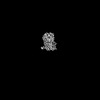
 Movie
Movie Controller


+ Open data

- Basic information
| Entry | Database: PDB / ID: 8zf4 | ||||||||||||||||||||||||
|---|---|---|---|---|---|---|---|---|---|---|---|---|---|---|---|---|---|---|---|---|---|---|---|---|---|
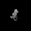
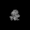
| Title | Cryo-EM structure of the receptor of xGPR4-Gs complex in pH6.2 | ||||||||||||||||||||||||
 Components Components | G-protein coupled receptor 4 | ||||||||||||||||||||||||
 Keywords Keywords | MEMBRANE PROTEIN / pH6.2 / xGPR4 / receptor / MEMBRANE PROTEIN/IMMUNE SYSTEM | ||||||||||||||||||||||||
| Function / homology |  Function and homology information Function and homology informationcellular response to acidic pH / G protein-coupled receptor activity / adenylate cyclase-activating G protein-coupled receptor signaling pathway / plasma membrane Similarity search - Function | ||||||||||||||||||||||||
| Biological species | |||||||||||||||||||||||||
| Method | ELECTRON MICROSCOPY / single particle reconstruction / cryo EM / Resolution: 3.05 Å | ||||||||||||||||||||||||
 Authors Authors | Rong, N.K. / Wen, X. / Yang, F. / Sun, J.P. | ||||||||||||||||||||||||
| Funding support |  China, 1items China, 1items
| ||||||||||||||||||||||||
 Citation Citation | Journal: Cell / Year: 2025 Title: Evolutionary study and structural basis of proton sensing by Mus GPR4 and Xenopus GPR4. Authors: Xin Wen / Pan Shang / Haidi Chen / Lulu Guo / Naikang Rong / Xiaoyu Jiang / Xuan Li / Junyan Liu / Gongming Yang / Jiacheng Zhang / Kongkai Zhu / Qingbiao Meng / Xuefei He / Zhihai Wang / ...Authors: Xin Wen / Pan Shang / Haidi Chen / Lulu Guo / Naikang Rong / Xiaoyu Jiang / Xuan Li / Junyan Liu / Gongming Yang / Jiacheng Zhang / Kongkai Zhu / Qingbiao Meng / Xuefei He / Zhihai Wang / Zili Liu / Haoran Cheng / Yilin Zheng / Bifei Zhang / Jiaojiao Pang / Zhaoqian Liu / Peng Xiao / Yuguo Chen / Lunxu Liu / Fengming Luo / Xiao Yu / Fan Yi / Pengju Zhang / Fan Yang / Cheng Deng / Jin-Peng Sun / Abstract: Animals have evolved pH-sensing membrane receptors, such as G-protein-coupled receptor 4 (GPR4), to monitor pH changes related to their physiology and generate adaptive reactions. However, the ...Animals have evolved pH-sensing membrane receptors, such as G-protein-coupled receptor 4 (GPR4), to monitor pH changes related to their physiology and generate adaptive reactions. However, the evolutionary trajectory and structural mechanism of proton sensing by GPR4 remain unresolved. Here, we observed a positive correlation between the optimal pH of GPR4 activity and the blood pH range across different species. By solving 7-cryoelectron microscopy (cryo-EM) structures of Xenopus tropicalis GPR4 (xtGPR4) and Mus musculus GPR4 (mmGPR4) under varying pH conditions, we identified that protonation of H and H enabled polar network establishment and tighter association between the extracellular loop 2 (ECL2) and 7 transmembrane (7TM) domain, as well as a conserved propagating path, which are common mechanisms underlying protonation-induced GPR4 activation across different species. Moreover, protonation of distinct extracellular H contributed to the more acidic optimal pH range of xtGPR4. Overall, our study revealed common and distinct mechanisms of proton sensing by GPR4, from a structural, functional, and evolutionary perspective. | ||||||||||||||||||||||||
| History |
|
- Structure visualization
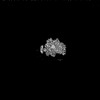
| Structure viewer | Molecule: MolmilJmol/JSmol |
|---|
- Downloads & links
-Download
| PDBx/mmCIF format | 8zf4.cif.gz | 60.5 KB | Display | PDBx/mmCIF format |
|---|---|---|---|---|
| PDB format | pdb8zf4.ent.gz | 41.3 KB | Display | PDB format |
| PDBx/mmJSON format | 8zf4.json.gz | Tree view | PDBx/mmJSON format | |
| Others |  Other downloads Other downloads |
-Validation report
| Arichive directory | https://data.pdbj.org/pub/pdb/validation_reports/zf/8zf4ftp://data.pdbj.org/pub/pdb/validation_reports/zf/8zf4 | HTTPS FTP |
|---|
-Related structure data
| Related structure data | 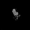 60050MC 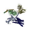 8zd1C 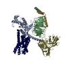 8zf6C 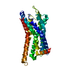 8zf7C 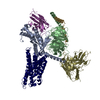 8zf9C 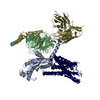 8zfaC 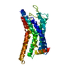 8zfbC 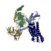 8zfcC 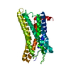 8zfdC 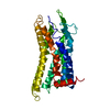 8zfeC 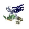 9jvgC 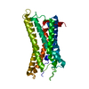 9jvhC 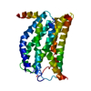 9jvmC M: map data used to model this data C: citing same article ( |
|---|---|
| Similar structure data |
-Links
 PDBj
PDBj


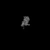
- Assembly
| Deposited unit | 
|
|---|---|
| 1 |
|
-Components
| #1: Protein | Mass: 38083.359 Da / Num. of mol.: 1 Source method: isolated from a genetically manipulated source Source: (gene. exp.) Gene: gpr4 / Production host:   Spodoptera frugiperda (fall armyworm) / References: UniProt: A0A6I8PUB9 Spodoptera frugiperda (fall armyworm) / References: UniProt: A0A6I8PUB9 |
|---|---|
| Has protein modification | Y |
-Experimental details
-Experiment
| Experiment | Method: ELECTRON MICROSCOPY |
|---|---|
| EM experiment | Aggregation state: PARTICLE / 3D reconstruction method: single particle reconstruction |
- Sample preparation
| Component | Name: Cryo-EM structure of the receptor of xGPR4-Gs complex in pH6.2 Type: COMPLEX / Entity ID: all / Source: RECOMBINANT |
|---|---|
| Source (natural) | Organism: |
| Source (recombinant) | Organism: Spodoptera frugiperda (fall armyworm) |
| Buffer solution | pH: 6.2 |
| Specimen | Embedding applied: NO / Shadowing applied: NO / Staining applied: NO / Vitrification applied: YES |
| Vitrification | Cryogen name: ETHANE |
- Electron microscopy imaging
| Experimental equipment |  Model: Titan Krios / Image courtesy: FEI Company |
|---|---|
| Microscopy | Model: TFS KRIOS |
| Electron gun | Electron source:  FIELD EMISSION GUN / Accelerating voltage: 300 kV / Illumination mode: FLOOD BEAM FIELD EMISSION GUN / Accelerating voltage: 300 kV / Illumination mode: FLOOD BEAM |
| Electron lens | Mode: DIFFRACTION / Nominal defocus max: 2000 nm / Nominal defocus min: 1000 nm |
| Image recording | Electron dose: 1.875 e/Å2 / Film or detector model: GATAN K3 (6k x 4k) |
- Processing
| EM software | Name: PHENIX / Category: model refinement | ||||||||||||||||||||||||
|---|---|---|---|---|---|---|---|---|---|---|---|---|---|---|---|---|---|---|---|---|---|---|---|---|---|
| CTF correction | Type: NONE | ||||||||||||||||||||||||
| 3D reconstruction | Resolution: 3.05 Å / Resolution method: FSC 0.143 CUT-OFF / Num. of particles: 640581 / Symmetry type: POINT | ||||||||||||||||||||||||
| Refine LS restraints |
|
