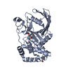
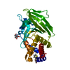
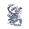
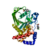
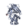
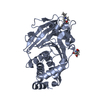
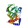
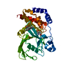
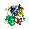
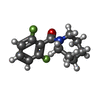
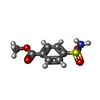
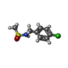
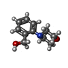
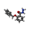
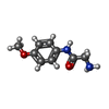
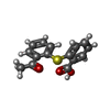
PDB-7gs7: PanDDA Analysis group deposition -- Crystal structure of PTP1B in complex with FMOPL000621a Method: X-RAY DIFFRACTION / Resolution: 1.66 Å
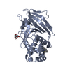
PDB-7gs8: PanDDA Analysis group deposition -- Crystal structure of PTP1B in complex with FMOPL000466a Method: X-RAY DIFFRACTION / Resolution: 1.67 Å
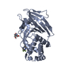
PDB-7gs9: PanDDA Analysis group deposition -- Crystal structure of PTP1B in complex with FMOPL000631a Method: X-RAY DIFFRACTION / Resolution: 1.96 Å
PDB-7gsa: PanDDA Analysis group deposition -- Crystal structure of PTP1B in complex with FMOPL000260a Method: X-RAY DIFFRACTION / Resolution: 1.72 Å
PDB-7gsb: PanDDA Analysis group deposition -- Crystal structure of PTP1B in complex with FMOPL000438a Method: X-RAY DIFFRACTION / Resolution: 1.72 Å
PDB-7gsc: PanDDA Analysis group deposition -- Crystal structure of PTP1B in complex with FMOPL000729a Method: X-RAY DIFFRACTION / Resolution: 1.69 Å
PDB-7gsd: PanDDA Analysis group deposition -- Crystal structure of PTP1B in complex with FMOPL000605a Method: X-RAY DIFFRACTION / Resolution: 1.8 Å
PDB-7gse: PanDDA Analysis group deposition -- Crystal structure of PTP1B in complex with FMOPL000383a Method: X-RAY DIFFRACTION / Resolution: 1.89 Å
PDB-7gsf: PanDDA Analysis group deposition -- Crystal structure of PTP1B in complex with FMOPL000421a Method: X-RAY DIFFRACTION / Resolution: 1.78 Å
PDB-7gsg: PanDDA Analysis group deposition -- Crystal structure of PTP1B in complex with FMOPL000316a Method: X-RAY DIFFRACTION / Resolution: 1.9 Å
PDB-7gsh: PanDDA Analysis group deposition -- Crystal structure of PTP1B in complex with FMOPL000530a Method: X-RAY DIFFRACTION / Resolution: 1.88 Å
PDB-7gsi: PanDDA Analysis group deposition -- Crystal structure of PTP1B in complex with XST00000046b Method: X-RAY DIFFRACTION / Resolution: 1.71 Å
PDB-7gsj: PanDDA Analysis group deposition -- Crystal structure of PTP1B in complex with FMOPL000543a Method: X-RAY DIFFRACTION / Resolution: 1.77 Å
PDB-7gsk: PanDDA Analysis group deposition -- Crystal structure of PTP1B in complex with FMOPL000279a Method: X-RAY DIFFRACTION / Resolution: 1.84 Å
PDB-7gsl: PanDDA Analysis group deposition -- Crystal structure of PTP1B in complex with FMSOA000274b Method: X-RAY DIFFRACTION / Resolution: 1.77 Å
PDB-7gsm: PanDDA Analysis group deposition -- Crystal structure of PTP1B in complex with XST00000437b Method: X-RAY DIFFRACTION / Resolution: 2.03 Å
PDB-7gsn: PanDDA Analysis group deposition -- Crystal structure of PTP1B in complex with XST00000519b Method: X-RAY DIFFRACTION / Resolution: 1.87 Å
PDB-7gso: PanDDA Analysis group deposition -- Crystal structure of PTP1B in complex with FMOMB000029a Method: X-RAY DIFFRACTION / Resolution: 1.72 Å
PDB-7gsq: PanDDA Analysis group deposition -- Crystal structure of PTP1B in complex with FMOMB000149a Method: X-RAY DIFFRACTION / Resolution: 1.73 Å
PDB-7gsr: PanDDA Analysis group deposition -- Crystal structure of PTP1B in complex with XST00000055b Method: X-RAY DIFFRACTION / Resolution: 1.69 Å
PDB-7gst: PanDDA Analysis group deposition -- Crystal structure of PTP1B in complex with FMOMB000056a Method: X-RAY DIFFRACTION / Resolution: 1.64 Å
PDB-7gsu: PanDDA Analysis group deposition -- Crystal structure of PTP1B in complex with FMOPL000382a Method: X-RAY DIFFRACTION / Resolution: 1.65 Å
PDB-7gsv: PanDDA Analysis group deposition -- Crystal structure of PTP1B in complex with FMSOA000830b Method: X-RAY DIFFRACTION / Resolution: 1.92 Å
PDB-7gsw: PanDDA Analysis group deposition -- Crystal structure of PTP1B in complex with XST00000422b Method: X-RAY DIFFRACTION / Resolution: 1.79 Å
PDB-7gsx: PanDDA Analysis group deposition -- Crystal structure of PTP1B in complex with FMSOA001440b Method: X-RAY DIFFRACTION / Resolution: 1.67 Å
PDB-7gsy: PanDDA Analysis group deposition -- Crystal structure of PTP1B in complex with FMSOA001175b Method: X-RAY DIFFRACTION / Resolution: 1.97 Å
PDB-7gsz: PanDDA Analysis group deposition -- Crystal structure of PTP1B in complex with FMSOA000686b Method: X-RAY DIFFRACTION / Resolution: 1.91 Å
PDB-7gt0: PanDDA Analysis group deposition -- Crystal structure of PTP1B in complex with FMOMB000275a Method: X-RAY DIFFRACTION / Resolution: 1.76 Å
PDB-7gt1: PanDDA Analysis group deposition -- Crystal structure of PTP1B in complex with FMOMB000209a Method: X-RAY DIFFRACTION / Resolution: 1.91 Å
PDB-7gt2: PanDDA Analysis group deposition -- Crystal structure of PTP1B in complex with XST00000752b Method: X-RAY DIFFRACTION / Resolution: 1.9 Å
PDB-7gt3: PanDDA Analysis group deposition -- Crystal structure of PTP1B in complex with FMOOA000527a Method: X-RAY DIFFRACTION / Resolution: 1.59 Å
PDB-7gt4: PanDDA Analysis group deposition -- Crystal structure of PTP1B in complex with FMOOA000528a Method: X-RAY DIFFRACTION / Resolution: 1.75 Å
PDB-7gt5: PanDDA Analysis group deposition -- Crystal structure of PTP1B in complex with FMOOA000529a Method: X-RAY DIFFRACTION / Resolution: 1.61 Å
PDB-7gt6: PanDDA Analysis group deposition -- Crystal structure of PTP1B in complex with FMOOA000530a Method: X-RAY DIFFRACTION / Resolution: 1.66 Å
PDB-7gt7: PanDDA Analysis group deposition -- Crystal structure of PTP1B in complex with FMSOA001181b Method: X-RAY DIFFRACTION / Resolution: 1.84 Å
PDB-7gt8: PanDDA Analysis group deposition -- Crystal structure of PTP1B in complex with FMSOA001439b Method: X-RAY DIFFRACTION / Resolution: 1.91 Å
PDB-7gt9: PanDDA Analysis group deposition -- Crystal structure of PTP1B in complex with FMSOA000463b Method: X-RAY DIFFRACTION / Resolution: 1.9 Å
PDB-7gta: PanDDA Analysis group deposition -- Crystal structure of PTP1B in complex with FMOMB000065a Method: X-RAY DIFFRACTION / Resolution: 2.06 Å
PDB-7gtb: PanDDA Analysis group deposition -- Crystal structure of PTP1B in complex with FMSOA000899b Method: X-RAY DIFFRACTION / Resolution: 1.86 Å
PDB-7gtc: PanDDA Analysis group deposition -- Crystal structure of PTP1B in complex with XST00001145b Method: X-RAY DIFFRACTION / Resolution: 1.92 Å
PDB-7gtd: PanDDA Analysis group deposition -- Crystal structure of PTP1B in complex with FMOMB000110a Method: X-RAY DIFFRACTION / Resolution: 1.91 Å
PDB-7gte: PanDDA Analysis group deposition -- Crystal structure of PTP1B in complex with XST00000646b Method: X-RAY DIFFRACTION / Resolution: 1.9 Å
PDB-7gtf: PanDDA Analysis group deposition -- Crystal structure of PTP1B in complex with XST00000754b Method: X-RAY DIFFRACTION / Resolution: 1.97 Å
PDB-7gtg: PanDDA Analysis group deposition -- Crystal structure of PTP1B in complex with FMOOA000684a Method: X-RAY DIFFRACTION / Resolution: 1.69 Å
PDB-7gth: PanDDA Analysis group deposition -- Crystal structure of PTP1B in complex with FMOOA000637a Method: X-RAY DIFFRACTION / Resolution: 1.66 Å
PDB-7gti: PanDDA Analysis group deposition -- Crystal structure of PTP1B in complex with FMOOA000571a Method: X-RAY DIFFRACTION / Resolution: 1.66 Å
PDB-7gtj: PanDDA Analysis group deposition -- Crystal structure of PTP1B in complex with XST00000280c Method: X-RAY DIFFRACTION / Resolution: 1.83 Å
PDB-7gtk: PanDDA Analysis group deposition -- Crystal structure of PTP1B in complex with FMOOA000552a Method: X-RAY DIFFRACTION / Resolution: 1.76 Å
PDB-7gtl: PanDDA analysis group deposition -- Crystal structure of PTP1B in complex with FMOOA000554a Method: X-RAY DIFFRACTION / Resolution: 1.83 Å
PDB-7gtm: PanDDA Analysis group deposition -- Crystal structure of PTP1B in complex with FMOOA000543a Method: X-RAY DIFFRACTION / Resolution: 1.72 Å
PDB-7gtn: PanDDA Analysis group deposition -- Crystal structure of PTP1B in complex with FMOOA000625a Method: X-RAY DIFFRACTION / Resolution: 1.66 Å
PDB-7gto: PanDDA Analysis group deposition -- Crystal structure of PTP1B in complex with FMOOA000602a Method: X-RAY DIFFRACTION / Resolution: 1.65 Å
PDB-7gtp: PanDDA Analysis group deposition -- Crystal structure of PTP1B in complex with FMOPL000688a Method: X-RAY DIFFRACTION / Resolution: 2.47 Å
PDB-7gtq: PanDDA Analysis group deposition -- Crystal structure of PTP1B in complex with FMOPL000311a Method: X-RAY DIFFRACTION / Resolution: 2.09 Å
PDB-7gtr: PanDDA Analysis group deposition -- Crystal structure of PTP1B in complex with FMOPL000587a Method: X-RAY DIFFRACTION / Resolution: 1.78 Å
PDB-7gts: PanDDA Analysis group deposition -- Crystal structure of PTP1B in complex with FMOPL000604a Method: X-RAY DIFFRACTION / Resolution: 1.8 Å
PDB-7gtt: PanDDA Analysis group deposition -- Crystal structure of PTP1B in complex with FMOPL000148a Method: X-RAY DIFFRACTION / Resolution: 1.93 Å
PDB-7gtu: PanDDA Analysis group deposition -- Crystal structure of PTP1B in complex with FMOMB000297a Method: X-RAY DIFFRACTION / Resolution: 2.08 Å
PDB-7gtv: PanDDA Analysis group deposition -- Crystal structure of PTP1B in complex with XST00000765c Method: X-RAY DIFFRACTION / Resolution: 1.72 Å
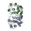
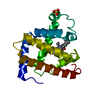
PDB-7gtw: PanDDA analysis group deposition of ground-state model of PTP1B, using pre-clustering, cluster1 Method: X-RAY DIFFRACTION / Resolution: 1.51 Å
PDB-7gtx: PanDDA analysis group deposition of ground-state model of PTP1B, using pre-clustering, cluster2 Method: X-RAY DIFFRACTION / Resolution: 1.51 Å
PDB-7gty: PanDDA analysis group deposition of ground-state model of PTP1B, using pre-clustering, cluster3 Method: X-RAY DIFFRACTION / Resolution: 1.54 Å
PDB-7gtz: PanDDA analysis group deposition of ground-state model of PTP1B, using pre-clustering, cluster5 Method: X-RAY DIFFRACTION / Resolution: 1.6 Å
PDB-7gu0: PanDDA analysis group deposition of ground-state model of PTP1B, using pre-clustering, cluster6 Method: X-RAY DIFFRACTION / Resolution: 1.66 Å
PDB-7gu1: PanDDA analysis group deposition of ground-state model of PTP1B, using pre-clustering, cluster7 Method: X-RAY DIFFRACTION / Resolution: 1.59 Å
PDB-7gu2: PanDDA analysis group deposition of ground-state model of PTP1B, using pre-clustering, cluster8 Method: X-RAY DIFFRACTION / Resolution: 1.8 Å
PDB-7gu3: PanDDA analysis group deposition of ground-state model of PTP1B, using pre-clustering, cluster9 Method: X-RAY DIFFRACTION / Resolution: 1.74 Å
PDB-7gu4: PanDDA analysis group deposition of ground-state model of PTP1B, using pre-clustering, cluster11 Method: X-RAY DIFFRACTION / Resolution: 1.59 Å
PDB-7gu5: PanDDA analysis group deposition of ground-state model of PTP1B, using pre-clustering, cluster12 Method: X-RAY DIFFRACTION / Resolution: 1.52 Å
PDB-7gu6: PanDDA analysis group deposition of ground-state model of PTP1B, using pre-clustering, cluster14 Method: X-RAY DIFFRACTION / Resolution: 1.56 Å
PDB-7gu7: PanDDA analysis group deposition of ground-state model of PTP1B, using pre-clustering, cluster15 Method: X-RAY DIFFRACTION / Resolution: 1.7 Å
PDB-7gu8: PanDDA analysis group deposition of ground-state model of PTP1B, using pre-clustering, cluster16 Method: X-RAY DIFFRACTION / Resolution: 1.59 Å
PDB-7gu9: PanDDA analysis group deposition of ground-state model of PTP1B, using pre-clustering, cluster17 Method: X-RAY DIFFRACTION / Resolution: 1.53 Å
PDB-7gua: PanDDA analysis group deposition of ground-state model of PTP1B, using pre-clustering, cluster18 Method: X-RAY DIFFRACTION / Resolution: 1.63 Å
PDB-7gub: PanDDA analysis group deposition of ground-state model of PTP1B, using pre-clustering, cluster19 Method: X-RAY DIFFRACTION / Resolution: 1.94 Å
PDB-7guc: PanDDA analysis group deposition of ground-state model of PTP1B, using pre-clustering, cluster20 Method: X-RAY DIFFRACTION / Resolution: 1.78 Å
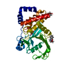
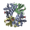
PDB-1abb: CONTROL OF PHOSPHORYLASE B CONFORMATION BY A MODIFIED COFACTOR: CRYSTALLOGRAPHIC STUDIES ON R-STATE GLYCOGEN PHOSPHORYLASE RECONSTITUTED WITH PYRIDOXAL 5'-DIPHOSPHATE
In the structure databanks used in Yorodumi, some data are registered as the other names, "COVID-19 virus" and "2019-nCoV". Here are the details of the virus and the list of structure data.
Jan 31, 2019. EMDB accession codes are about to change! (news from PDBe EMDB page)
EMDB accession codes are about to change! (news from PDBe EMDB page)
The allocation of 4 digits for EMDB accession codes will soon come to an end. Whilst these codes will remain in use, new EMDB accession codes will include an additional digit and will expand incrementally as the available range of codes is exhausted. The current 4-digit format prefixed with “EMD-” (i.e. EMD-XXXX) will advance to a 5-digit format (i.e. EMD-XXXXX), and so on. It is currently estimated that the 4-digit codes will be depleted around Spring 2019, at which point the 5-digit format will come into force.
The EM Navigator/Yorodumi systems omit the EMD- prefix.
Related info.:Q: What is EMD? / ID/Accession-code notation in Yorodumi/EM Navigator
 Movie
Movie Controller
 Structure viewers
 About Yorodumi Papers


 Authors
 External links


 Keywords
 homo sapiens (human)