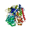
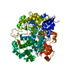
 Movie
Movie Controller


+ Open data

- Basic information
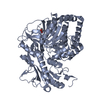
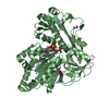
| Entry | Database: PDB / ID: 1aa6 | ||||||
|---|---|---|---|---|---|---|---|
| Title | REDUCED FORM OF FORMATE DEHYDROGENASE H FROM E. COLI | ||||||
 Components Components | FORMATE DEHYDROGENASE H | ||||||
 Keywords Keywords | OXIDOREDUCTASE / SELENIUM / SELENOCYSTEINE / SECYS / MOLYBDENUM / MOLYBDOPTERIN / MPT / MOLYBDOPTERIN GUANINE DINUCLEOTIDE / MGD / IRON SULFUR CLUSTER / FE4S4 / FORMATE / DEHYDROGENASE / ANAEROBIC | ||||||
| Function / homology |  Function and homology information Function and homology informationformate dehydrogenase (hydrogenase) / formate oxidation / oxidoreductase activity, acting on the aldehyde or oxo group of donors / formate dehydrogenase complex / glucose catabolic process / formate dehydrogenase (NAD+) activity / anaerobic electron transport chain / urate catabolic process / molybdopterin cofactor binding / anaerobic respiration ...formate dehydrogenase (hydrogenase) / formate oxidation / oxidoreductase activity, acting on the aldehyde or oxo group of donors / formate dehydrogenase complex / glucose catabolic process / formate dehydrogenase (NAD+) activity / anaerobic electron transport chain / urate catabolic process / molybdopterin cofactor binding / anaerobic respiration / respiratory electron transport chain / 4 iron, 4 sulfur cluster binding / membrane / metal ion binding / cytosol Similarity search - Function | ||||||
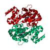
| Biological species |  | ||||||
| Method |  X-RAY DIFFRACTION / SYNCHROTRON / MIR, MAD / Resolution: 2.3 Å X-RAY DIFFRACTION / SYNCHROTRON / MIR, MAD / Resolution: 2.3 Å | ||||||
 Authors Authors | Sun, P.D. / Boyington, J.C. | ||||||
 Citation Citation | Journal: Science / Year: 1997 Title: Crystal structure of formate dehydrogenase H: catalysis involving Mo, molybdopterin, selenocysteine, and an Fe4S4 cluster. Authors: Boyington, J.C. / Gladyshev, V.N. / Khangulov, S.V. / Stadtman, T.C. / Sun, P.D. #1: Journal: J.Biol.Chem. / Year: 1996Title: Characterization of Crystalline Formate Dehydrogenase H from Escherichia Coli. Stabilization, Epr Spectroscopy, and Preliminary Crystallographic Analysis Authors: Gladyshev, V.N. / Boyington, J.C. / Khangulov, S.V. / Grahame, D.A. / Stadtman, T.C. / Sun, P.D. | ||||||
| History |
|
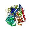
- Structure visualization
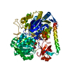
| Structure viewer | Molecule: MolmilJmol/JSmol |
|---|
- Downloads & links
-Download
| PDBx/mmCIF format | 1aa6.cif.gz | 152.7 KB | Display | PDBx/mmCIF format |
|---|---|---|---|---|
| PDB format | pdb1aa6.ent.gz | 118.6 KB | Display | PDB format |
| PDBx/mmJSON format | 1aa6.json.gz | Tree view | PDBx/mmJSON format | |
| Others |  Other downloads Other downloads |
-Validation report
| Arichive directory | https://data.pdbj.org/pub/pdb/validation_reports/aa/1aa6ftp://data.pdbj.org/pub/pdb/validation_reports/aa/1aa6 | HTTPS FTP |
|---|
-Related structure data

-Links
 PDBj
PDBj


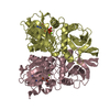
- Assembly
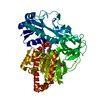
| Deposited unit | 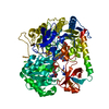
| ||||||||
|---|---|---|---|---|---|---|---|---|---|
| 1 |
| ||||||||
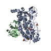
| Unit cell |
|
-Components
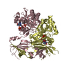
| #1: Protein | Mass: 79465.703 Da / Num. of mol.: 1 Source method: isolated from a genetically manipulated source Details: REDUCED FORM (MO(IV),FE4S4(RED)) OF FDH-H / Source: (gene. exp.) | ||||
|---|---|---|---|---|---|
| #2: Chemical | ChemComp-SF4 /   Mass: 351.640 Da / Num. of mol.: 1 / Source method: obtained synthetically / Formula: Fe4S4 Mass: 351.640 Da / Num. of mol.: 1 / Source method: obtained synthetically / Formula: Fe4S4 | ||||
| #3: Chemical | 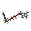  Mass: 740.557 Da / Num. of mol.: 2 / Source method: obtained synthetically / Formula: C20H26N10O13P2S2 Mass: 740.557 Da / Num. of mol.: 2 / Source method: obtained synthetically / Formula: C20H26N10O13P2S2#4: Chemical | ChemComp-4MO / |   Mass: 95.940 Da / Num. of mol.: 1 / Source method: obtained synthetically / Formula: Mo Mass: 95.940 Da / Num. of mol.: 1 / Source method: obtained synthetically / Formula: Mo#5: Water | ChemComp-HOH / |  Mass: 18.015 Da / Num. of mol.: 83 / Source method: isolated from a natural source / Formula: H2O Mass: 18.015 Da / Num. of mol.: 83 / Source method: isolated from a natural source / Formula: H2O |
-Experimental details
-Experiment
| Experiment | Method: X-RAY DIFFRACTION / Number of used crystals: 1 |
|---|
- Sample preparation
| Crystal | Density Matthews: 2.77 Å3/Da / Density % sol: 56 % | ||||||||||||||||||||||||||||||
|---|---|---|---|---|---|---|---|---|---|---|---|---|---|---|---|---|---|---|---|---|---|---|---|---|---|---|---|---|---|---|---|
| Crystal grow | Method: vapor diffusion - hanging drop in an anaerobic atmosphere pH: 7.5 Details: FDH-H WAS CRYSTALLIZED BY HANGING DROP VAPOR DIFFUSION IN AN ANAEROBIC ATMOSPHERE IN THE PRESENCE OF 1.5 M AMMONIUM SULFATE, 1% PEG 400, 20 MM SODIUM FORMATE AND 100 MM HEPES/NAOH AT PH 7.5, ...Details: FDH-H WAS CRYSTALLIZED BY HANGING DROP VAPOR DIFFUSION IN AN ANAEROBIC ATMOSPHERE IN THE PRESENCE OF 1.5 M AMMONIUM SULFATE, 1% PEG 400, 20 MM SODIUM FORMATE AND 100 MM HEPES/NAOH AT PH 7.5, vapor diffusion - hanging drop in an anaerobic atmosphere | ||||||||||||||||||||||||||||||
| Crystal grow | *PLUS Method: vapor diffusion, hanging dropDetails: drop solution was mixed with an equal volume of reservoir solution | ||||||||||||||||||||||||||||||
| Components of the solutions | *PLUS
|
-Data collection
| Diffraction | Mean temperature: 100 K |
|---|---|
| Diffraction source | Source: SYNCHROTRON / Site: NSLS  / Beamline: X4A / Wavelength: 1.0402 / Beamline: X4A / Wavelength: 1.0402 |
| Detector | Type: FUJI / Detector: IMAGE PLATE / Date: Jun 1, 1996 |
| Radiation | Monochromatic (M) / Laue (L): M / Scattering type: x-ray |
| Radiation wavelength | Wavelength: 1.0402 Å / Relative weight: 1 |
| Reflection | Resolution: 2.3→50 Å / Num. obs: 36025 / % possible obs: 88 % / Observed criterion σ(I): 1 / Redundancy: 4.7 % / Rmerge(I) obs: 0.085 / Net I/σ(I): 26.1 |
| Reflection shell | Resolution: 2.3→2.38 Å / Rmerge(I) obs: 0.19 / Mean I/σ(I) obs: 8.4 / % possible all: 65 |
| Reflection | *PLUS Num. measured all: 169244 |
| Reflection shell | *PLUS % possible obs: 65 % |
- Processing
| Software |
| ||||||||||||||||||||||||||||||||||||||||||||||||||||||||||||||||||||||||||||||||
|---|---|---|---|---|---|---|---|---|---|---|---|---|---|---|---|---|---|---|---|---|---|---|---|---|---|---|---|---|---|---|---|---|---|---|---|---|---|---|---|---|---|---|---|---|---|---|---|---|---|---|---|---|---|---|---|---|---|---|---|---|---|---|---|---|---|---|---|---|---|---|---|---|---|---|---|---|---|---|---|---|---|
| Refinement | Method to determine structure: MIR, MAD / Resolution: 2.3→6 Å / Rfactor Rfree error: 0.007 / Data cutoff high absF: 100000 / Data cutoff low absF: 0.1 / Isotropic thermal model: RESTRAINED / Cross valid method: THROUGHOUT / σ(F): 2
| ||||||||||||||||||||||||||||||||||||||||||||||||||||||||||||||||||||||||||||||||
| Displacement parameters | Biso mean: 28.4 Å2 | ||||||||||||||||||||||||||||||||||||||||||||||||||||||||||||||||||||||||||||||||
| Refinement step | Cycle: LAST / Resolution: 2.3→6 Å
| ||||||||||||||||||||||||||||||||||||||||||||||||||||||||||||||||||||||||||||||||
| Refine LS restraints |
| ||||||||||||||||||||||||||||||||||||||||||||||||||||||||||||||||||||||||||||||||
| LS refinement shell | Resolution: 2.3→2.38 Å / Rfactor Rfree error: 0.027 / Total num. of bins used: 10
| ||||||||||||||||||||||||||||||||||||||||||||||||||||||||||||||||||||||||||||||||
| Xplor file |
| ||||||||||||||||||||||||||||||||||||||||||||||||||||||||||||||||||||||||||||||||
| Software | *PLUS Name: X-PLOR / Version: 3.1 / Classification: refinement | ||||||||||||||||||||||||||||||||||||||||||||||||||||||||||||||||||||||||||||||||
| Refinement | *PLUS | ||||||||||||||||||||||||||||||||||||||||||||||||||||||||||||||||||||||||||||||||
| Solvent computation | *PLUS | ||||||||||||||||||||||||||||||||||||||||||||||||||||||||||||||||||||||||||||||||
| Displacement parameters | *PLUS | ||||||||||||||||||||||||||||||||||||||||||||||||||||||||||||||||||||||||||||||||
| Refine LS restraints | *PLUS
| ||||||||||||||||||||||||||||||||||||||||||||||||||||||||||||||||||||||||||||||||
| LS refinement shell | *PLUS Rfactor obs: 0.269 |
