 Movie
Movie Controller
 Structure viewers
 About Yorodumi Papers


+Search query
-Structure paper
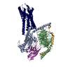
| Title | Molecular basis of proton sensing by G protein-coupled receptors. |
|---|---|
| Journal, issue, pages | Cell, Vol. 188, Issue 3, Page 671-687.e20, Year 2025 |
| Publish date | Feb 6, 2025 |
 Authors Authors | Matthew K Howard / Nicholas Hoppe / Xi-Ping Huang / Darko Mitrovic / Christian B Billesbølle / Christian B Macdonald / Eshan Mehrotra / Patrick Rockefeller Grimes / Donovan D Trinidad / Lucie Delemotte / Justin G English / Willow Coyote-Maestas / Aashish Manglik /   |
| PubMed Abstract | Three proton-sensing G protein-coupled receptors (GPCRs)-GPR4, GPR65, and GPR68-respond to extracellular pH to regulate diverse physiology. How protons activate these receptors is poorly understood. ...Three proton-sensing G protein-coupled receptors (GPCRs)-GPR4, GPR65, and GPR68-respond to extracellular pH to regulate diverse physiology. How protons activate these receptors is poorly understood. We determined cryogenic-electron microscopy (cryo-EM) structures of each receptor to understand the spatial arrangement of proton-sensing residues. Using deep mutational scanning (DMS), we determined the functional importance of every residue in GPR68 activation by generating ∼9,500 mutants and measuring their effects on signaling and surface expression. Constant-pH molecular dynamics simulations provided insights into the conformational landscape and protonation patterns of key residues. This unbiased approach revealed that, unlike other proton-sensitive channels and receptors, no single site is critical for proton recognition. Instead, a network of titratable residues extends from the extracellular surface to the transmembrane region, converging on canonical motifs to activate proton-sensing GPCRs. Our approach integrating structure, simulations, and unbiased functional interrogation provides a framework for understanding GPCR signaling complexity. |
 External links External links | Cell / PubMed:39753132 / PubMed Central |
| Methods | EM (single particle) |
| Resolution | 2.8 - 3.0 Å |
| Structure data |  EMDB-44548: Human proton sensing receptor GPR65 in complex with miniGs - focused map of 7TM EMDB-44549, PDB-9bhl: EMDB-44550, PDB-9bhm: EMDB-44560, PDB-9bi6: 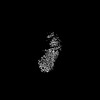 EMDB-44561: Human proton sensing receptor GPR68 in complex with miniGsq - focused map of 7TM 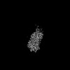 EMDB-44596: Human proton sensing receptor GPR4 in complex with miniGs - focused map of 7TM EMDB-44597, PDB-9bip: |
| Chemicals |  ChemComp-Y01: |
| Source |
|
 Keywords Keywords | SIGNALING PROTEIN / Receptor / Proton-sensor / G protein |


 homo sapiens (human)
