 Movie
Movie Controller

[English] 日本語
 Yorodumi
- EMDB-44548: Human proton sensing receptor GPR65 in complex with miniGs - focu... -
+ Open data

- Basic information
| Entry |  | |||||||||
|---|---|---|---|---|---|---|---|---|---|---|
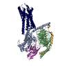
| Title | Human proton sensing receptor GPR65 in complex with miniGs - focused map of 7TM | |||||||||
 Map data Map data | ||||||||||
 Sample Sample |
| |||||||||
 Keywords Keywords | Receptor / Proton-sensor / G protein / SIGNALING PROTEIN | |||||||||
| Biological species |  Homo sapiens (human) / Homo sapiens (human) /  | |||||||||
| Method | single particle reconstruction / cryo EM / Resolution: 3.0 Å | |||||||||
 Authors Authors | Howard MK / Hoppe N / Huang XP / Macdonald CB / Mehrotra E / Rockefeller Grimes P / Zahm AM / Trinidad DD / English J / Coyote-Maestas W / Manglik A | |||||||||
| Funding support |  United States, 1 items United States, 1 items
| |||||||||
 Citation Citation | Journal: Cell / Year: 2025 Title: Molecular basis of proton sensing by G protein-coupled receptors. Authors: Matthew K Howard / Nicholas Hoppe / Xi-Ping Huang / Darko Mitrovic / Christian B Billesbølle / Christian B Macdonald / Eshan Mehrotra / Patrick Rockefeller Grimes / Donovan D Trinidad / ...Authors: Matthew K Howard / Nicholas Hoppe / Xi-Ping Huang / Darko Mitrovic / Christian B Billesbølle / Christian B Macdonald / Eshan Mehrotra / Patrick Rockefeller Grimes / Donovan D Trinidad / Lucie Delemotte / Justin G English / Willow Coyote-Maestas / Aashish Manglik /  Abstract: Three proton-sensing G protein-coupled receptors (GPCRs)-GPR4, GPR65, and GPR68-respond to extracellular pH to regulate diverse physiology. How protons activate these receptors is poorly understood. ...Three proton-sensing G protein-coupled receptors (GPCRs)-GPR4, GPR65, and GPR68-respond to extracellular pH to regulate diverse physiology. How protons activate these receptors is poorly understood. We determined cryogenic-electron microscopy (cryo-EM) structures of each receptor to understand the spatial arrangement of proton-sensing residues. Using deep mutational scanning (DMS), we determined the functional importance of every residue in GPR68 activation by generating ∼9,500 mutants and measuring their effects on signaling and surface expression. Constant-pH molecular dynamics simulations provided insights into the conformational landscape and protonation patterns of key residues. This unbiased approach revealed that, unlike other proton-sensitive channels and receptors, no single site is critical for proton recognition. Instead, a network of titratable residues extends from the extracellular surface to the transmembrane region, converging on canonical motifs to activate proton-sensing GPCRs. Our approach integrating structure, simulations, and unbiased functional interrogation provides a framework for understanding GPCR signaling complexity. | |||||||||
| History |
|
- Structure visualization
| Supplemental images |
|---|

- Downloads & links
-EMDB archive
| Map data | emd_44548.map.gz | 95.2 MB |  EMDB map data format EMDB map data format | |
|---|---|---|---|---|
| Header (meta data) | emd-44548-v30.xmlemd-44548.xml | 16.9 KB 16.9 KB | Display Display | EMDB header |
| Images |  emd_44548.png emd_44548.png | 53 KB | ||
| Filedesc metadata | emd-44548.cif.gz | 4.4 KB | ||
| Others | emd_44548_additional_1.map.gzemd_44548_half_map_1.map.gzemd_44548_half_map_2.map.gz | 94.8 MB 22.2 MB 22.2 MB | ||
| Archive directory |  http://ftp.pdbj.org/pub/emdb/structures/EMD-44548ftp://ftp.pdbj.org/pub/emdb/structures/EMD-44548 http://ftp.pdbj.org/pub/emdb/structures/EMD-44548ftp://ftp.pdbj.org/pub/emdb/structures/EMD-44548 | HTTPS FTP |
-Related structure data


-Links
| EMDB pages | EMDB (EBI/PDBe) / EMDataResource |
|---|
-Map
| File | Download / File: emd_44548.map.gz / Format: CCP4 / Size: 103 MB / Type: IMAGE STORED AS FLOATING POINT NUMBER (4 BYTES) | ||||||||||||||||||||||||||||||||||||
|---|---|---|---|---|---|---|---|---|---|---|---|---|---|---|---|---|---|---|---|---|---|---|---|---|---|---|---|---|---|---|---|---|---|---|---|---|---|
| Projections & slices | Image control
Images are generated by Spider. | ||||||||||||||||||||||||||||||||||||
| Voxel size | X=Y=Z: 0.835 Å | ||||||||||||||||||||||||||||||||||||
| Density |
| ||||||||||||||||||||||||||||||||||||
| Symmetry | Space group: 1 | ||||||||||||||||||||||||||||||||||||
| Details | EMDB XML:
|
 Z (Sec.)
 Y (Row.)
 X (Col.)


-Supplemental data
-Additional map: #1
| File | emd_44548_additional_1.map | ||||||||||||
|---|---|---|---|---|---|---|---|---|---|---|---|---|---|
| Projections & Slices |
| ||||||||||||
| Density Histograms |


-Half map: #1
| File | emd_44548_half_map_1.map | ||||||||||||
|---|---|---|---|---|---|---|---|---|---|---|---|---|---|
| Projections & Slices |
| ||||||||||||
| Density Histograms |


-Half map: #2
| File | emd_44548_half_map_2.map | ||||||||||||
|---|---|---|---|---|---|---|---|---|---|---|---|---|---|
| Projections & Slices |
| ||||||||||||
| Density Histograms |


- Sample components
-Entire : GPR65 receptor in complex with miniGs
| Entire | Name: GPR65 receptor in complex with miniGs |
|---|---|
| Components |
|
-Supramolecule #1: GPR65 receptor in complex with miniGs
| Supramolecule | Name: GPR65 receptor in complex with miniGs / type: complex / ID: 1 / Parent: 0 |
|---|
-Supramolecule #2: GPR65 receptor
| Supramolecule | Name: GPR65 receptor / type: complex / ID: 2 / Parent: 1 |
|---|---|
| Source (natural) | Organism: Homo sapiens (human) |
-Supramolecule #3: miniGs (alpha and beta subunits)
| Supramolecule | Name: miniGs (alpha and beta subunits) / type: complex / ID: 3 / Parent: 1 |
|---|---|
| Source (natural) | Organism: Homo sapiens (human) |
-Supramolecule #4: Nanobody 35
| Supramolecule | Name: Nanobody 35 / type: complex / ID: 4 / Parent: 1 |
|---|---|
| Source (natural) | Organism: |
-Experimental details
-Structure determination
| Method | cryo EM |
|---|---|
 Processing Processing | single particle reconstruction |
| Aggregation state | particle |
-Sample preparation
| Buffer | pH: 6 |
|---|---|
| Vitrification | Cryogen name: ETHANE |
- Electron microscopy
| Microscope | FEI TITAN KRIOS |
|---|---|
| Image recording | Film or detector model: GATAN K3 BIOQUANTUM (6k x 4k) / Average electron dose: 46.0 e/Å2 |
| Electron beam | Acceleration voltage: 300 kV / Electron source:  FIELD EMISSION GUN FIELD EMISSION GUN |
| Electron optics | Illumination mode: FLOOD BEAM / Imaging mode: BRIGHT FIELD / Nominal defocus max: 2.1 µm / Nominal defocus min: 1.0 µm |
| Experimental equipment |  Model: Titan Krios / Image courtesy: FEI Company |
-Image processing
| Startup model | Type of model: NONE |
|---|---|
| Final reconstruction | Resolution.type: BY AUTHOR / Resolution: 3.0 Å / Resolution method: FSC 0.143 CUT-OFF / Number images used: 411592 |
| Initial angle assignment | Type: MAXIMUM LIKELIHOOD |
| Final angle assignment | Type: MAXIMUM LIKELIHOOD |
