 Movie
Movie Controller


+ Open data

- Basic information
| Entry |  | |||||||||||||||||||||
|---|---|---|---|---|---|---|---|---|---|---|---|---|---|---|---|---|---|---|---|---|---|---|
| Title | Human proton sensing receptor GPR4 in complex with miniGs | |||||||||||||||||||||
 Map data Map data | Human proton sensing receptor GPR4 in complex with miniGs | |||||||||||||||||||||
 Sample Sample |
| |||||||||||||||||||||
 Keywords Keywords | Receptor / Proton-sensor / G protein / SIGNALING PROTEIN | |||||||||||||||||||||
| Function / homology |  Function and homology information Function and homology informationregulation of vascular permeability / Class A/1 (Rhodopsin-like receptors) / response to acidic pH / positive regulation of Rho protein signal transduction / regulation of cell adhesion / cellular response to acidic pH / G protein-coupled receptor activity / Olfactory Signaling Pathway / Activation of the phototransduction cascade / G protein-coupled acetylcholine receptor signaling pathway ...regulation of vascular permeability / Class A/1 (Rhodopsin-like receptors) / response to acidic pH / positive regulation of Rho protein signal transduction / regulation of cell adhesion / cellular response to acidic pH / G protein-coupled receptor activity / Olfactory Signaling Pathway / Activation of the phototransduction cascade / G protein-coupled acetylcholine receptor signaling pathway / G beta:gamma signalling through PLC beta / Presynaptic function of Kainate receptors / Thromboxane signalling through TP receptor / positive regulation of inflammatory response / Activation of G protein gated Potassium channels / Inhibition of voltage gated Ca2+ channels via Gbeta/gamma subunits / G-protein activation / Glucagon signaling in metabolic regulation / G beta:gamma signalling through CDC42 / Prostacyclin signalling through prostacyclin receptor / Synthesis, secretion, and inactivation of Glucagon-like Peptide-1 (GLP-1) / G beta:gamma signalling through BTK / photoreceptor disc membrane / ADP signalling through P2Y purinoceptor 12 / Glucagon-type ligand receptors / Sensory perception of sweet, bitter, and umami (glutamate) taste / Adrenaline,noradrenaline inhibits insulin secretion / Vasopressin regulates renal water homeostasis via Aquaporins / Glucagon-like Peptide-1 (GLP1) regulates insulin secretion / G alpha (z) signalling events / cellular response to catecholamine stimulus / ADP signalling through P2Y purinoceptor 1 / G beta:gamma signalling through PI3Kgamma / ADORA2B mediated anti-inflammatory cytokines production / adenylate cyclase-activating dopamine receptor signaling pathway / Cooperation of PDCL (PhLP1) and TRiC/CCT in G-protein beta folding / GPER1 signaling / cellular response to prostaglandin E stimulus / heterotrimeric G-protein complex / Inactivation, recovery and regulation of the phototransduction cascade / G alpha (12/13) signalling events / G-protein beta-subunit binding / extracellular vesicle / sensory perception of taste / Thrombin signalling through proteinase activated receptors (PARs) / signaling receptor complex adaptor activity / adenylate cyclase-activating G protein-coupled receptor signaling pathway / retina development in camera-type eye / fibroblast proliferation / GTPase binding / Ca2+ pathway / High laminar flow shear stress activates signaling by PIEZO1 and PECAM1:CDH5:KDR in endothelial cells / G alpha (i) signalling events / G alpha (s) signalling events / G alpha (q) signalling events / phospholipase C-activating G protein-coupled receptor signaling pathway / Ras protein signal transduction / cell population proliferation / Extra-nuclear estrogen signaling / G protein-coupled receptor signaling pathway / lysosomal membrane / GTPase activity / synapse / protein-containing complex binding / signal transduction / extracellular exosome / membrane / plasma membrane / cytosol / cytoplasm Similarity search - Function | |||||||||||||||||||||
| Biological species |  Homo sapiens (human) / Homo sapiens (human) /  | |||||||||||||||||||||
| Method | single particle reconstruction / cryo EM / Resolution: 2.8 Å | |||||||||||||||||||||
 Authors Authors | Howard MK / Hoppe N / Huang XP / Macdonald CB / Mehrotra E / Rockefeller Grimes P / Zahm AM / Trinidad DD / English J / Coyote-Maestas W / Manglik A | |||||||||||||||||||||
| Funding support |  United States, 6 items United States, 6 items
| |||||||||||||||||||||
 Citation Citation | Journal: Cell / Year: 2025 Title: Molecular basis of proton sensing by G protein-coupled receptors. Authors: Matthew K Howard / Nicholas Hoppe / Xi-Ping Huang / Darko Mitrovic / Christian B Billesbølle / Christian B Macdonald / Eshan Mehrotra / Patrick Rockefeller Grimes / Donovan D Trinidad / ...Authors: Matthew K Howard / Nicholas Hoppe / Xi-Ping Huang / Darko Mitrovic / Christian B Billesbølle / Christian B Macdonald / Eshan Mehrotra / Patrick Rockefeller Grimes / Donovan D Trinidad / Lucie Delemotte / Justin G English / Willow Coyote-Maestas / Aashish Manglik /  Abstract: Three proton-sensing G protein-coupled receptors (GPCRs)-GPR4, GPR65, and GPR68-respond to extracellular pH to regulate diverse physiology. How protons activate these receptors is poorly understood. ...Three proton-sensing G protein-coupled receptors (GPCRs)-GPR4, GPR65, and GPR68-respond to extracellular pH to regulate diverse physiology. How protons activate these receptors is poorly understood. We determined cryogenic-electron microscopy (cryo-EM) structures of each receptor to understand the spatial arrangement of proton-sensing residues. Using deep mutational scanning (DMS), we determined the functional importance of every residue in GPR68 activation by generating ∼9,500 mutants and measuring their effects on signaling and surface expression. Constant-pH molecular dynamics simulations provided insights into the conformational landscape and protonation patterns of key residues. This unbiased approach revealed that, unlike other proton-sensitive channels and receptors, no single site is critical for proton recognition. Instead, a network of titratable residues extends from the extracellular surface to the transmembrane region, converging on canonical motifs to activate proton-sensing GPCRs. Our approach integrating structure, simulations, and unbiased functional interrogation provides a framework for understanding GPCR signaling complexity. | |||||||||||||||||||||
| History |
|
- Structure visualization
| Supplemental images |
|---|

- Downloads & links
-EMDB archive
| Map data | emd_44597.map.gz | 85.9 MB | EMDB map data format | |
|---|---|---|---|---|
| Header (meta data) | emd-44597-v30.xmlemd-44597.xml | 22.2 KB 22.2 KB | Display Display | EMDB header |
| Images |  emd_44597.png emd_44597.png | 101.1 KB | ||
| Filedesc metadata | emd-44597.cif.gz | 6.8 KB | ||
| Others | emd_44597_additional_1.map.gzemd_44597_half_map_1.map.gzemd_44597_half_map_2.map.gz | 45.1 MB 84.6 MB 84.6 MB | ||
| Archive directory |  http://ftp.pdbj.org/pub/emdb/structures/EMD-44597ftp://ftp.pdbj.org/pub/emdb/structures/EMD-44597 http://ftp.pdbj.org/pub/emdb/structures/EMD-44597ftp://ftp.pdbj.org/pub/emdb/structures/EMD-44597 | HTTPS FTP |
-Related structure data
| Related structure data | 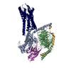 9bipMC  9bhlC  9bhmC  9bi6C C: citing same article ( M: atomic model generated by this map |
|---|---|
| Similar structure data |


-Links
| EMDB pages | EMDB (EBI/PDBe) / EMDataResource |
|---|---|
| Related items in Molecule of the Month |


-Map
| File | Download / File: emd_44597.map.gz / Format: CCP4 / Size: 91.1 MB / Type: IMAGE STORED AS FLOATING POINT NUMBER (4 BYTES) | ||||||||||||||||||||||||||||||||||||
|---|---|---|---|---|---|---|---|---|---|---|---|---|---|---|---|---|---|---|---|---|---|---|---|---|---|---|---|---|---|---|---|---|---|---|---|---|---|
| Annotation | Human proton sensing receptor GPR4 in complex with miniGs | ||||||||||||||||||||||||||||||||||||
| Projections & slices | Image control
Images are generated by Spider. | ||||||||||||||||||||||||||||||||||||
| Voxel size | X=Y=Z: 0.86 Å | ||||||||||||||||||||||||||||||||||||
| Density |
| ||||||||||||||||||||||||||||||||||||
| Symmetry | Space group: 1 | ||||||||||||||||||||||||||||||||||||
| Details | EMDB XML:
|
 Z (Sec.)
 Y (Row.)
 X (Col.)


-Supplemental data
-Additional map: Additional map
| File | emd_44597_additional_1.map | ||||||||||||
|---|---|---|---|---|---|---|---|---|---|---|---|---|---|
| Annotation | Additional map | ||||||||||||
| Projections & Slices |
| ||||||||||||
| Density Histograms |


-Half map: Half Map B
| File | emd_44597_half_map_1.map | ||||||||||||
|---|---|---|---|---|---|---|---|---|---|---|---|---|---|
| Annotation | Half Map B | ||||||||||||
| Projections & Slices |
| ||||||||||||
| Density Histograms |


-Half map: Half Map A
| File | emd_44597_half_map_2.map | ||||||||||||
|---|---|---|---|---|---|---|---|---|---|---|---|---|---|
| Annotation | Half Map A | ||||||||||||
| Projections & Slices |
| ||||||||||||
| Density Histograms |


- Sample components
-Entire : Complex of GPR4 bound to Gs heterotrimer
| Entire | Name: Complex of GPR4 bound to Gs heterotrimer |
|---|---|
| Components |
|
-Supramolecule #1: Complex of GPR4 bound to Gs heterotrimer
| Supramolecule | Name: Complex of GPR4 bound to Gs heterotrimer / type: complex / ID: 1 / Parent: 0 / Macromolecule list: all |
|---|---|
| Source (natural) | Organism: Homo sapiens (human) |
-Macromolecule #1: miniGs
| Macromolecule | Name: miniGs / type: protein_or_peptide / ID: 1 / Number of copies: 1 / Enantiomer: LEVO |
|---|---|
| Source (natural) | Organism: Homo sapiens (human) |
| Molecular weight | Theoretical: 30.137025 KDa |
| Recombinant expression | Organism: Homo sapiens (human) |
| Sequence | String: GGSLEVLFQG PSGNSKTEDQ RNEEKAQREA NKKIEKQLQK DKQVYRATHR LLLLGADNSG KSTIVKQMRI LHGGSGGSGG TSGIFETKF QVDKVNFHMF DVGGQRDERR KWIQCFNDVT AIIFVVDSSD YNRLQEALNL FKSIWNNRWL RTISVILFLN K QDLLAEKV ...String: GGSLEVLFQG PSGNSKTEDQ RNEEKAQREA NKKIEKQLQK DKQVYRATHR LLLLGADNSG KSTIVKQMRI LHGGSGGSGG TSGIFETKF QVDKVNFHMF DVGGQRDERR KWIQCFNDVT AIIFVVDSSD YNRLQEALNL FKSIWNNRWL RTISVILFLN K QDLLAEKV LAGKSKIEDY FPEFARYTTP EDATPEPGED PRVTRAKYFI RDEFLRISTA SGDGRHYCYP HFTCAVDTEN AR RIFNDCR DIIQRMHLRQ YELL |
-Macromolecule #2: Guanine nucleotide-binding protein G(I)/G(S)/G(T) subunit beta-1
| Macromolecule | Name: Guanine nucleotide-binding protein G(I)/G(S)/G(T) subunit beta-1 type: protein_or_peptide / ID: 2 / Number of copies: 1 / Enantiomer: LEVO |
|---|---|
| Source (natural) | Organism: Homo sapiens (human) |
| Molecular weight | Theoretical: 40.786566 KDa |
| Recombinant expression | Organism:  Trichoplusia ni (cabbage looper) Trichoplusia ni (cabbage looper) |
| Sequence | String: MHHHHHHLEV LFQGPEDQVD PRLIDGKGSS GSELDQLRQE AEQLKNQIRD ARKACADATL SQITNNIDPV GRIQMRTRRT LRGHLAKIY AMHWGTDSRL LVSASQDGKL IIWDSYTTNK VHAIPLRSSW VMTCAYAPSG NYVACGGLDN ICSIYNLKTR E GNVRVSRE ...String: MHHHHHHLEV LFQGPEDQVD PRLIDGKGSS GSELDQLRQE AEQLKNQIRD ARKACADATL SQITNNIDPV GRIQMRTRRT LRGHLAKIY AMHWGTDSRL LVSASQDGKL IIWDSYTTNK VHAIPLRSSW VMTCAYAPSG NYVACGGLDN ICSIYNLKTR E GNVRVSRE LAGHTGYLSC CRFLDDNQIV TSSGDTTCAL WDIETGQQTT TFTGHTGDVM SLSLAPDTRL FVSGACDASA KL WDVREGM CRQTFTGHES DINAICFFPN GNAFATGSDD ATCRLFDLRA DQELMTYSHD NIICGITSVS FSKSGRLLLA GYD DFNCNV WDALKADRAG VLAGHDNRVS CLGVTDDGMA VATGSWDSFL KIWN UniProtKB: Guanine nucleotide-binding protein G(I)/G(S)/G(T) subunit beta-1 |
-Macromolecule #3: Guanine nucleotide-binding protein G(I)/G(S)/G(O) subunit gamma-2
| Macromolecule | Name: Guanine nucleotide-binding protein G(I)/G(S)/G(O) subunit gamma-2 type: protein_or_peptide / ID: 3 / Number of copies: 1 / Enantiomer: LEVO |
|---|---|
| Source (natural) | Organism: Homo sapiens (human) |
| Molecular weight | Theoretical: 7.56375 KDa |
| Recombinant expression | Organism: Trichoplusia ni (cabbage looper) |
| Sequence | String: MASNNTASIA QARKLVEQLK MEANIDRIKV SKAAADLMAY CEAHAKEDPL LTPVPASENP FREKKFFC UniProtKB: Guanine nucleotide-binding protein G(I)/G(S)/G(O) subunit gamma-2 |
-Macromolecule #4: Nb35
| Macromolecule | Name: Nb35 / type: protein_or_peptide / ID: 4 / Number of copies: 1 / Enantiomer: LEVO |
|---|---|
| Source (natural) | Organism: |
| Molecular weight | Theoretical: 15.398067 KDa |
| Recombinant expression | Organism:  |
| Sequence | String: QVQLQESGGG LVQPGGSLRL SCAASGFTFS NYKMNWVRQA PGKGLEWVSD ISQSGASISY TGSVKGRFTI SRDNAKNTLY LQMNSLKPE DTAVYYCARC PAPFTRDCFD VTSTTYAYRG QGTQVTVSSG SEDQVDPRLI DGK |
-Macromolecule #5: G-protein coupled receptor 4
| Macromolecule | Name: G-protein coupled receptor 4 / type: protein_or_peptide / ID: 5 / Number of copies: 1 / Enantiomer: LEVO |
|---|---|
| Source (natural) | Organism: Homo sapiens (human) |
| Molecular weight | Theoretical: 42.280852 KDa |
| Recombinant expression | Organism: Homo sapiens (human) |
| Sequence | String: DYKDDDDASI DMGNHTWEGC HVDSRVDHLF PPSLYIFVIG VGLPTNCLAL WAAYRQVQQR NELGVYLMNL SIADLLYICT LPLWVDYFL HHDNWIHGPG SCKLFGFIFY TNIYISIAFL CCISVDRYLA VAHPLRFARL RRVKTAVAVS SVVWATELGA N SAPLFHDE ...String: DYKDDDDASI DMGNHTWEGC HVDSRVDHLF PPSLYIFVIG VGLPTNCLAL WAAYRQVQQR NELGVYLMNL SIADLLYICT LPLWVDYFL HHDNWIHGPG SCKLFGFIFY TNIYISIAFL CCISVDRYLA VAHPLRFARL RRVKTAVAVS SVVWATELGA N SAPLFHDE LFRDRYNHTF CFEKFPMEGW VAWMNLYRVF VGFLFPWALM LLSYRGILRA VRGSVSTERQ EKAKIKRLAL SL IAIVLVC FAPYHVLLLS RSAIYLGRPW DCGFEERVFS AYHSSLAFTS LNCVADPILY CLVNEGARSD VAKALHNLLR FLA SDKPQE MANASLTLET PLTSKRNSTA KAMTGSWAAT PPSQGDQVQL KMLPPAQ UniProtKB: G protein-coupled receptor 4 |
-Experimental details
-Structure determination
| Method | cryo EM |
|---|---|
 Processing Processing | single particle reconstruction |
| Aggregation state | particle |
-Sample preparation
| Buffer | pH: 6 |
|---|---|
| Vitrification | Cryogen name: ETHANE |
- Electron microscopy
| Microscope | FEI TITAN KRIOS |
|---|---|
| Image recording | Film or detector model: GATAN K3 BIOQUANTUM (6k x 4k) / Average electron dose: 60.0 e/Å2 |
| Electron beam | Acceleration voltage: 300 kV / Electron source:  FIELD EMISSION GUN FIELD EMISSION GUN |
| Electron optics | Illumination mode: FLOOD BEAM / Imaging mode: BRIGHT FIELD / Nominal defocus max: 2.1 µm / Nominal defocus min: 1.0 µm |
| Experimental equipment |  Model: Titan Krios / Image courtesy: FEI Company |
-Image processing
| Startup model | Type of model: NONE |
|---|---|
| Final reconstruction | Resolution.type: BY AUTHOR / Resolution: 2.8 Å / Resolution method: FSC 0.143 CUT-OFF / Number images used: 439296 |
| Initial angle assignment | Type: MAXIMUM LIKELIHOOD |
| Final angle assignment | Type: MAXIMUM LIKELIHOOD |
