 Movie
Movie Controller


+ Open data

- Basic information
| Entry | Database: PDB / ID: 9bhl | |||||||||||||||||||||||||||||||||||||||
|---|---|---|---|---|---|---|---|---|---|---|---|---|---|---|---|---|---|---|---|---|---|---|---|---|---|---|---|---|---|---|---|---|---|---|---|---|---|---|---|---|
| Title | Human proton sensing receptor GPR65 in complex with miniGs | |||||||||||||||||||||||||||||||||||||||
 Components Components |
| |||||||||||||||||||||||||||||||||||||||
 Keywords Keywords | SIGNALING PROTEIN / Receptor / Proton-sensor / G protein | |||||||||||||||||||||||||||||||||||||||
| Function / homology |  Function and homology information Function and homology informationpositive regulation of interleukin-22 production / positive regulation of T-helper cell differentiation / Class A/1 (Rhodopsin-like receptors) / response to acidic pH / adenylate cyclase-activating G protein-coupled bile acid receptor signaling pathway / adenylate cyclase-activating serotonin receptor signaling pathway / regulation of skeletal muscle contraction / hair follicle placode formation / PKA activation in glucagon signalling / developmental growth ...positive regulation of interleukin-22 production / positive regulation of T-helper cell differentiation / Class A/1 (Rhodopsin-like receptors) / response to acidic pH / adenylate cyclase-activating G protein-coupled bile acid receptor signaling pathway / adenylate cyclase-activating serotonin receptor signaling pathway / regulation of skeletal muscle contraction / hair follicle placode formation / PKA activation in glucagon signalling / developmental growth / intracellular transport / D1 dopamine receptor binding / renal water homeostasis / vascular endothelial cell response to laminar fluid shear stress / Hedgehog 'off' state / activation of adenylate cyclase activity / positive regulation of stress fiber assembly / adenylate cyclase-activating adrenergic receptor signaling pathway / cellular response to acidic pH / cellular response to glucagon stimulus / intracellular glucose homeostasis / positive regulation of insulin secretion involved in cellular response to glucose stimulus / adenylate cyclase activator activity / trans-Golgi network membrane / negative regulation of inflammatory response to antigenic stimulus / response to prostaglandin E / bone development / platelet aggregation / cognition / G protein-coupled receptor activity / positive regulation of insulin secretion / G-protein beta/gamma-subunit complex binding / Olfactory Signaling Pathway / Activation of the phototransduction cascade / G protein-coupled acetylcholine receptor signaling pathway / sensory perception of smell / G beta:gamma signalling through PLC beta / Presynaptic function of Kainate receptors / Thromboxane signalling through TP receptor / Activation of G protein gated Potassium channels / Inhibition of voltage gated Ca2+ channels via Gbeta/gamma subunits / G-protein activation / Glucagon signaling in metabolic regulation / G beta:gamma signalling through CDC42 / Prostacyclin signalling through prostacyclin receptor / Synthesis, secretion, and inactivation of Glucagon-like Peptide-1 (GLP-1) / G beta:gamma signalling through BTK / photoreceptor disc membrane / ADP signalling through P2Y purinoceptor 12 / Glucagon-type ligand receptors / Sensory perception of sweet, bitter, and umami (glutamate) taste / Adrenaline,noradrenaline inhibits insulin secretion / Vasopressin regulates renal water homeostasis via Aquaporins / Glucagon-like Peptide-1 (GLP1) regulates insulin secretion / G alpha (z) signalling events / late endosome membrane / cellular response to catecholamine stimulus / ADP signalling through P2Y purinoceptor 1 / G beta:gamma signalling through PI3Kgamma / ADORA2B mediated anti-inflammatory cytokines production / adenylate cyclase-activating dopamine receptor signaling pathway / Cooperation of PDCL (PhLP1) and TRiC/CCT in G-protein beta folding / cellular response to prostaglandin E stimulus / positive regulation of cold-induced thermogenesis / GPER1 signaling / heterotrimeric G-protein complex / Inactivation, recovery and regulation of the phototransduction cascade / G alpha (12/13) signalling events / extracellular vesicle / Thrombin signalling through proteinase activated receptors (PARs) / signaling receptor complex adaptor activity / adenylate cyclase-activating G protein-coupled receptor signaling pathway / GTPase binding / G protein activity / early endosome membrane / Ca2+ pathway / High laminar flow shear stress activates signaling by PIEZO1 and PECAM1:CDH5:KDR in endothelial cells / G alpha (i) signalling events / G alpha (s) signalling events / G alpha (q) signalling events / Hydrolases; Acting on acid anhydrides; Acting on GTP to facilitate cellular and subcellular movement / Ras protein signal transduction / Extra-nuclear estrogen signaling / immune response / G protein-coupled receptor signaling pathway / lysosomal membrane / GTPase activity / apoptotic process / synapse / GTP binding / protein-containing complex binding / signal transduction / extracellular exosome / membrane / metal ion binding / plasma membrane / cytosol / cytoplasm Similarity search - Function | |||||||||||||||||||||||||||||||||||||||
| Biological species |  Homo sapiens (human) Homo sapiens (human) | |||||||||||||||||||||||||||||||||||||||
| Method | ELECTRON MICROSCOPY / single particle reconstruction / cryo EM / Resolution: 2.8 Å | |||||||||||||||||||||||||||||||||||||||
 Authors Authors | Howard, M.K. / Hoppe, N. / Huang, X.P. / Macdonald, C.B. / Mehrotra, E. / Rockefeller Grimes, P. / Zahm, A.M. / Trinidad, D.D. / English, J. / Coyote-Maestas, W. / Manglik, A. | |||||||||||||||||||||||||||||||||||||||
| Funding support |  United States, 6items United States, 6items
| |||||||||||||||||||||||||||||||||||||||
 Citation Citation | Journal: Cell / Year: 2025 Title: Molecular basis of proton sensing by G protein-coupled receptors. Authors: Matthew K Howard / Nicholas Hoppe / Xi-Ping Huang / Darko Mitrovic / Christian B Billesbølle / Christian B Macdonald / Eshan Mehrotra / Patrick Rockefeller Grimes / Donovan D Trinidad / ...Authors: Matthew K Howard / Nicholas Hoppe / Xi-Ping Huang / Darko Mitrovic / Christian B Billesbølle / Christian B Macdonald / Eshan Mehrotra / Patrick Rockefeller Grimes / Donovan D Trinidad / Lucie Delemotte / Justin G English / Willow Coyote-Maestas / Aashish Manglik /  Abstract: Three proton-sensing G protein-coupled receptors (GPCRs)-GPR4, GPR65, and GPR68-respond to extracellular pH to regulate diverse physiology. How protons activate these receptors is poorly understood. ...Three proton-sensing G protein-coupled receptors (GPCRs)-GPR4, GPR65, and GPR68-respond to extracellular pH to regulate diverse physiology. How protons activate these receptors is poorly understood. We determined cryogenic-electron microscopy (cryo-EM) structures of each receptor to understand the spatial arrangement of proton-sensing residues. Using deep mutational scanning (DMS), we determined the functional importance of every residue in GPR68 activation by generating ∼9,500 mutants and measuring their effects on signaling and surface expression. Constant-pH molecular dynamics simulations provided insights into the conformational landscape and protonation patterns of key residues. This unbiased approach revealed that, unlike other proton-sensitive channels and receptors, no single site is critical for proton recognition. Instead, a network of titratable residues extends from the extracellular surface to the transmembrane region, converging on canonical motifs to activate proton-sensing GPCRs. Our approach integrating structure, simulations, and unbiased functional interrogation provides a framework for understanding GPCR signaling complexity. | |||||||||||||||||||||||||||||||||||||||
| History |
|
- Structure visualization
| Structure viewer | Molecule: MolmilJmol/JSmol |
|---|
- Downloads & links
-Download
| PDBx/mmCIF format | 9bhl.cif.gz | 419.2 KB | Display | PDBx/mmCIF format |
|---|---|---|---|---|
| PDB format | pdb9bhl.ent.gz | 342.4 KB | Display | PDB format |
| PDBx/mmJSON format | 9bhl.json.gz | Tree view | PDBx/mmJSON format | |
| Others |  Other downloads Other downloads |
-Validation report
| Arichive directory | https://data.pdbj.org/pub/pdb/validation_reports/bh/9bhlftp://data.pdbj.org/pub/pdb/validation_reports/bh/9bhl | HTTPS FTP |
|---|
-Related structure data
| Related structure data |  44549MC  9bhmC  9bi6C 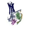 9bipC C: citing same article ( M: map data used to model this data |
|---|---|
| Similar structure data |


-Links
 PDBj
PDBj


- Assembly
| Deposited unit | 
|
|---|---|
| 1 |
|
-Components
| #1: Protein | Mass: 30137.025 Da / Num. of mol.: 1 Mutation: G49D,E50N,residues 65-203 replaced with GGSGGSGG,A249D,S252D,residues 254-263 deleted,I372A,V375I Source method: isolated from a genetically manipulated source Details: miniGs / Source: (gene. exp.) Homo sapiens (human) / Gene: GNAS, GNAS1, GSP / Production host: Homo sapiens (human) / References: UniProt: P63092 |
|---|---|
| #2: Protein | Mass: 40857.641 Da / Num. of mol.: 1 Source method: isolated from a genetically manipulated source Source: (gene. exp.) Homo sapiens (human) / Gene: GNB1 / Production host: Homo sapiens (human) / References: UniProt: P62873 |
| #3: Antibody | Mass: 15398.067 Da / Num. of mol.: 1 Source method: isolated from a genetically manipulated source Source: (gene. exp.)  |
| #4: Protein | Mass: 40627.215 Da / Num. of mol.: 1 Source method: isolated from a genetically manipulated source Source: (gene. exp.) Homo sapiens (human) / Gene: GPR65, TDAG8 / Production host: Homo sapiens (human) / References: UniProt: Q8IYL9 |
| Has protein modification | Y |
-Experimental details
-Experiment
| Experiment | Method: ELECTRON MICROSCOPY |
|---|---|
| EM experiment | Aggregation state: PARTICLE / 3D reconstruction method: single particle reconstruction |
- Sample preparation
| Component | Name: Complex of GPR65 bound to Gs heterotrimer / Type: COMPLEX / Entity ID: all / Source: MULTIPLE SOURCES |
|---|---|
| Molecular weight | Experimental value: NO |
| Buffer solution | pH: 6 |
| Specimen | Embedding applied: NO / Shadowing applied: NO / Staining applied: NO / Vitrification applied: YES |
| Vitrification | Cryogen name: ETHANE |
- Electron microscopy imaging
| Experimental equipment |  Model: Titan Krios / Image courtesy: FEI Company |
|---|---|
| Microscopy | Model: FEI TITAN KRIOS |
| Electron gun | Electron source:  FIELD EMISSION GUN / Accelerating voltage: 300 kV / Illumination mode: FLOOD BEAM FIELD EMISSION GUN / Accelerating voltage: 300 kV / Illumination mode: FLOOD BEAM |
| Electron lens | Mode: BRIGHT FIELD / Nominal defocus max: 2100 nm / Nominal defocus min: 1000 nm |
| Image recording | Electron dose: 46 e/Å2 / Film or detector model: GATAN K3 BIOQUANTUM (6k x 4k) |
- Processing
| EM software | Name: PHENIX / Category: model refinement | ||||||||||||||||||||||||
|---|---|---|---|---|---|---|---|---|---|---|---|---|---|---|---|---|---|---|---|---|---|---|---|---|---|
| CTF correction | Type: PHASE FLIPPING AND AMPLITUDE CORRECTION | ||||||||||||||||||||||||
| 3D reconstruction | Resolution: 2.8 Å / Resolution method: FSC 0.143 CUT-OFF / Num. of particles: 411592 / Symmetry type: POINT | ||||||||||||||||||||||||
| Refine LS restraints |
|
