[English] 日本語
Yorodumi Papers
- Database of articles cited by EMDB/PDB/SASBDB data -

+
Search query

Keywords
Structure methods
Author
Journal
IF

-
Structure paper

TitleStructural basis for differential inhibition of eukaryotic ribosomes by tigecycline.
Journal, issue, pagesNat Commun, Vol. 15, Issue 1, Page 5481, Year 2024
Publish dateJun 28, 2024
AuthorsXiang Li / Mengjiao Wang / Timo Denk / Robert Buschauer / Yi Li / Roland Beckmann / Jingdong Cheng /
PubMed AbstractTigecycline is widely used for treating complicated bacterial infections for which there are no effective drugs. It inhibits bacterial protein translation by blocking the ribosomal A-site. However, ...Tigecycline is widely used for treating complicated bacterial infections for which there are no effective drugs. It inhibits bacterial protein translation by blocking the ribosomal A-site. However, even though it is also cytotoxic for human cells, the molecular mechanism of its inhibition remains unclear. Here, we present cryo-EM structures of tigecycline-bound human mitochondrial 55S, 39S, cytoplasmic 80S and yeast cytoplasmic 80S ribosomes. We find that at clinically relevant concentrations, tigecycline effectively targets human 55S mitoribosomes, potentially, by hindering A-site tRNA accommodation and by blocking the peptidyl transfer center. In contrast, tigecycline does not bind to human 80S ribosomes under physiological concentrations. However, at high tigecycline concentrations, in addition to blocking the A-site, both human and yeast 80S ribosomes bind tigecycline at another conserved binding site restricting the movement of the L1 stalk. In conclusion, the observed distinct binding properties of tigecycline may guide new pathways for drug design and therapy.
External linksNat Commun / PubMed:38942792 / PubMed Central
MethodsEM (single particle)
Resolution2.0 - 3.6 Å
Structure data

EMDB-36836, PDB-8k2a:
Cryo-EM structure of the human 55S mitoribosome with Tigecycline
Method: EM (single particle) / Resolution: 2.9 Å

EMDB-36837, PDB-8k2b:
Cryo-EM structure of the human 39S mitoribosome with Tigecycline
Method: EM (single particle) / Resolution: 3.4 Å

EMDB-36838, PDB-8k2c:
Cryo-EM structure of the human 80S ribosome with Tigecycline
Method: EM (single particle) / Resolution: 2.4 Å

EMDB-36839, PDB-8k2d:
Cryo-EM structure of the yeast 80S ribosome with tigecycline, eEF2, Stm1 and eIF5A
Method: EM (single particle) / Resolution: 3.2 Å

EMDB-36945, PDB-8k82:
Cryo-EM structure of the yeast 80S ribosome with tigecycline, Not5 and P-site tRNA
Method: EM (single particle) / Resolution: 3.0 Å

EMDB-38629, PDB-8xsx:
Cryo-EM structure of the human 80S ribosome with Tigecycline, E-tRNA, SERBP1 and eEF2
Method: EM (single particle) / Resolution: 2.4 Å

EMDB-38630, PDB-8xsy:
Cryo-EM structure of the human 80S ribosome with Tigecycline, e-tRNA and CCDC124 (40S head Swivelled)
Method: EM (single particle) / Resolution: 3.0 Å

EMDB-38631, PDB-8xsz:
Cryo-EM structure of the human 80S ribosome with Tigecycline, E-tRNA and P-tRNA
Method: EM (single particle) / Resolution: 3.2 Å

EMDB-38632, PDB-8xt0:
Cryo-EM structure of the human 55S mitoribosome with 5um Tigecycline
Method: EM (single particle) / Resolution: 3.2 Å

EMDB-38633, PDB-8xt1:
Cryo-EM structure of the human 39S mitoribosome with 5uM Tigecycline
Method: EM (single particle) / Resolution: 3.1 Å

EMDB-38634, PDB-8xt2:
Cryo-EM structure of the human 55S mitoribosome with 10uM Tigecycline
Method: EM (single particle) / Resolution: 3.3 Å

EMDB-38635, PDB-8xt3:
Cryo-EM structure of the human 39S mitoribosome with 10uM Tigecycline
Method: EM (single particle) / Resolution: 3.1 Å

EMDB-38636: Cryo-EM structure of the human 55S mitoribosome with 5uM Tigecycline (focusing on 39S ribosome)
Method: EM (single particle) / Resolution: 3.1 Å

EMDB-38637: Cryo-EM structure of the human 55S mitoribosome with 5uM Tigecycline (focusing on 28S ribosome head)
Method: EM (single particle) / Resolution: 3.4 Å

EMDB-38638: Cryo-EM structure of the human 55S mitoribosome with 10uM Tigecycline (focusing on 39S ribosome)
Method: EM (single particle) / Resolution: 3.2 Å

EMDB-38639: Cryo-EM structure of the human 55S mitoribosome with 10uM Tigecycline (focusing on 28S ribosome head)
Method: EM (single particle) / Resolution: 3.6 Å

EMDB-39455, PDB-8yoo:
Cryo-EM structure of the human 80S ribosome with 100 um Tigecycline
Method: EM (single particle) / Resolution: 2.0 Å

EMDB-39456, PDB-8yop:
Cryo-EM structure of the human 80S ribosome with 4 um Tigecycline
Method: EM (single particle) / Resolution: 2.2 Å

Chemicals

ChemComp-MG:
Unknown entry

ChemComp-T1C:
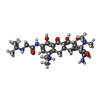
TIGECYCLINE / medication, antibiotic*YM

ChemComp-ZN:
Unknown entry

ChemComp-GDP:
GUANOSINE-5'-DIPHOSPHATE / GDP, energy-carrying molecule*YM

ChemComp-PNS:
4'-PHOSPHOPANTETHEINE

ChemComp-SPD:
SPERMIDINE

ChemComp-ADP:
ADENOSINE-5'-DIPHOSPHATE / ADP, energy-carrying molecule*YM

Source
  • homo sapiens (human)
  • saccharomyces cerevisiae (brewer's yeast)
KeywordsRIBOSOME / 55S mitoribosome / Tigecycline / antibiotic / 39S mitoribosome / 80S ribosome / eEF2 / SERBP1 / CCDC124

+
About Yorodumi Papers

-
News

-
Feb 9, 2022. New format data for meta-information of EMDB entries

New format data for meta-information of EMDB entries

  • Version 3 of the EMDB header file is now the official format.
  • The previous official version 1.9 will be removed from the archive.

Related info.:EMDB header

External links:wwPDB to switch to version 3 of the EMDB data model

-
Aug 12, 2020. Covid-19 info

Covid-19 info

URL: https://pdbj.org/emnavi/covid19.php

New page: Covid-19 featured information page in EM Navigator.

Related info.:Covid-19 info / Mar 5, 2020. Novel coronavirus structure data

+
Mar 5, 2020. Novel coronavirus structure data

Novel coronavirus structure data

Related info.:Yorodumi Speices / Aug 12, 2020. Covid-19 info

External links:COVID-19 featured content - PDBj / Molecule of the Month (242):Coronavirus Proteases

+
Jan 31, 2019. EMDB accession codes are about to change! (news from PDBe EMDB page)

EMDB accession codes are about to change! (news from PDBe EMDB page)

  • The allocation of 4 digits for EMDB accession codes will soon come to an end. Whilst these codes will remain in use, new EMDB accession codes will include an additional digit and will expand incrementally as the available range of codes is exhausted. The current 4-digit format prefixed with “EMD-” (i.e. EMD-XXXX) will advance to a 5-digit format (i.e. EMD-XXXXX), and so on. It is currently estimated that the 4-digit codes will be depleted around Spring 2019, at which point the 5-digit format will come into force.
  • The EM Navigator/Yorodumi systems omit the EMD- prefix.

Related info.:Q: What is EMD? / ID/Accession-code notation in Yorodumi/EM Navigator

External links:EMDB Accession Codes are Changing Soon! / Contact to PDBj

+
Jul 12, 2017. Major update of PDB

Major update of PDB

  • wwPDB released updated PDB data conforming to the new PDBx/mmCIF dictionary.
  • This is a major update changing the version number from 4 to 5, and with Remediation, in which all the entries are updated.
  • In this update, many items about electron microscopy experimental information are reorganized (e.g. em_software).
  • Now, EM Navigator and Yorodumi are based on the updated data.

External links:wwPDB Remediation / Enriched Model Files Conforming to OneDep Data Standards Now Available in the PDB FTP Archive

-
Yorodumi Papers

Database of articles cited by EMDB/PDB/SASBDB data

  • Database of articles cited by EMDB, PDB, and SASBDB entries
  • Using PubMed data

Related info.:EMDB / PDB / SASBDB / Yorodumi / EMN Papers / Changes in new EM Navigator and Yorodumi

Read more