Matteo Bianchi / Hannah L Turner / Bartek Nogal / Christopher A Cottrell / David Oyen / Matthias Pauthner / Raiza Bastidas / Rebecca Nedellec / Laura E McCoy / Ian A Wilson / Dennis R Burton / Andrew B Ward / Lars Hangartner /
PubMed Abstract
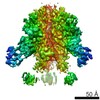
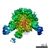
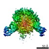
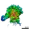
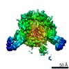
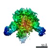
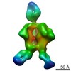
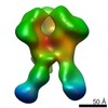
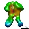
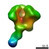
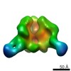
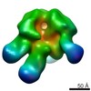
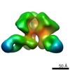
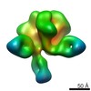
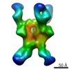
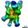
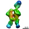
Characterizing polyclonal antibody responses via currently available methods is inherently complex and difficult. Mapping epitopes in an immune response is typically incomplete, which creates a ...Characterizing polyclonal antibody responses via currently available methods is inherently complex and difficult. Mapping epitopes in an immune response is typically incomplete, which creates a barrier to fully understanding the humoral response to antigens and hinders rational vaccine design efforts. Here, we describe a method of characterizing polyclonal responses by using electron microscopy, and we applied this method to the immunization of rabbits with an HIV-1 envelope glycoprotein vaccine candidate, BG505 SOSIP.664. We detected known epitopes within the polyclonal sera and revealed how antibody responses evolved during the prime-boosting strategy to ultimately result in a neutralizing antibody response. We uncovered previously unidentified epitopes, including an epitope proximal to one recognized by human broadly neutralizing antibodies as well as potentially distracting non-neutralizing epitopes. Our method provides an efficient and semiquantitative map of epitopes that are targeted in a polyclonal antibody response and should be of widespread utility in vaccine and infection studies.
EMDB-7552: CryoEM map of BG505 SOSIP.664 in complex with post boost 1 serum from rabbit 3417 Method: EM (single particle) / Resolution: 4.7 Å
EMDB-7553: CryoEM map of BG505 SOSIP.664 in complex with post boost 1 serum from rabbit 3417 Method: EM (single particle) / Resolution: 4.7 Å
EMDB-7554: CryoEM map of BG505 SOSIP.664 in complex with post boost 1 serum from rabbit 3417 Method: EM (single particle) / Resolution: 4.7 Å
EMDB-7555: CryoEM map of BG505 SOSIP.664 in complex with post boost 1 serum from rabbit 3417 Method: EM (single particle) / Resolution: 4.7 Å
EMDB-7556: CryoEM map of BG505 SOSIP.664 in complex with post boost 1 serum from rabbit 3417 Method: EM (single particle) / Resolution: 4.7 Å
EMDB-7557: CryoEM map of BG505 SOSIP.664 in complex with post boost 1 serum from rabbit 3417 Method: EM (single particle) / Resolution: 4.7 Å
EMDB-7570: Negative stain EM map of BG505 SOSIP.644 in complex with PG9 and 12N Fabs Method: EM (single particle) / Resolution: 17.36 Å
EMDB-7887: Negative Stain EM map of polyclonal serum in complex with BG505 SOSIP.664 from rabbit 3418 at post boost 2 Method: EM (single particle) / Resolution: 20.0 Å
EMDB-7888: Negative Stain EM map of polyclonal serum in complex with BG505 SOSIP.664 from rabbit 3418 at post boost 2. Method: EM (single particle) / Resolution: 20.0 Å
EMDB-7889: Negative Stain EM map of polyclonal serum in complex with BG505 SOSIP.664 from rabbit 3419 at post boost 2. Method: EM (single particle) / Resolution: 20.0 Å
EMDB-7890: Negative Stain EM map of polyclonal serum in complex with BG505 SOSIP.664 from rabbit 3419 at post boost 2. Method: EM (single particle) / Resolution: 20.0 Å
EMDB-7891: Negative Stain EM map of polyclonal serum in complex with BG505 SOSIP.664 from rabbit 3417 at post boost 2. Method: EM (single particle) / Resolution: 20.0 Å
EMDB-7892: Negative Stain EM map of polyclonal serum in complex with BG505 SOSIP.664 from rabbit 3417 at post boost 2. Method: EM (single particle) / Resolution: 20.0 Å
EMDB-7893: Negative Stain EM map of polyclonal serum in complex with BG505 SOSIP.664 from rabbit 3417 at post boost 2. Method: EM (single particle) / Resolution: 20.0 Å
EMDB-7894: Negative Stain EM map of polyclonal serum in complex with BG505 SOSIP.664 from rabbit 3420 at post boost 2. Method: EM (single particle) / Resolution: 20.0 Å
EMDB-7895: Negative Stain EM map of polyclonal serum in complex with BG505 MD39 CPG9 from rabbit 3417 at post boost 3 Method: EM (single particle) / Resolution: 20.0 Å
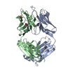
EMDB-7896, PDB-6did: HIV Env BG505 SOSIP with polyclonal Fabs from immunized rabbit #3417 post-boost#1 Method: EM (single particle) / Resolution: 4.71 Å
EMDB-7903: Negative stain EM map of BG505 SOSIP.644 in complex with PGT121 and 12N Fabs Method: EM (single particle) / Resolution: 17.36 Å
EMDB-7904: Negative stain EM map of BG505 SOSIP.644 in complex with PGT121 and serum from rabbit 3417 PB1 Method: EM (single particle) / Resolution: 18.45 Å
EMDB-7906: Negative stain EM map of BG505 SOSIP.644 in complex with PG9 Fab and serum from rabbit 3417 PB1 Method: EM (single particle) / Resolution: 18.45 Å
PDB-6cjk: Anti HIV Fab 10A Method: X-RAY DIFFRACTION / Resolution: 1.795 Å
In the structure databanks used in Yorodumi, some data are registered as the other names, "COVID-19 virus" and "2019-nCoV". Here are the details of the virus and the list of structure data.
Jan 31, 2019. EMDB accession codes are about to change! (news from PDBe EMDB page)
EMDB accession codes are about to change! (news from PDBe EMDB page)
The allocation of 4 digits for EMDB accession codes will soon come to an end. Whilst these codes will remain in use, new EMDB accession codes will include an additional digit and will expand incrementally as the available range of codes is exhausted. The current 4-digit format prefixed with “EMD-” (i.e. EMD-XXXX) will advance to a 5-digit format (i.e. EMD-XXXXX), and so on. It is currently estimated that the 4-digit codes will be depleted around Spring 2019, at which point the 5-digit format will come into force.
The EM Navigator/Yorodumi systems omit the EMD- prefix.
Related info.:Q: What is EMD? / ID/Accession-code notation in Yorodumi/EM Navigator
 Movie
Movie Controller
 Structure viewers
 About Yorodumi Papers


 Authors

 External links
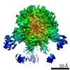


 Keywords
 IMMUNE SYSTEM /