[English] 日本語
Yorodumi Papers
- Database of articles cited by EMDB/PDB/SASBDB data -

+
Search query

Keywords
Structure methods
Author
Journal
IF

-
Structure paper

TitleTargeting a Pleckstrin Homology Domain with a Lysine-Reactive Covalent Binder.
Journal, issue, pagesJ. Med. Chem., Year 2026
Publish dateJan 7, 2020 (structure data deposition date)
AuthorsWest, R.M. / Bizga Nicolescu, R.C. / Brear, P. / Wagstaff, J. / Blaszczyk, B.K. / Deingruber, T. / Sanders, M.G. / Perez-Areales, F.J. / Spring, D.R. / Hyvonen, M.
External linksJ. Med. Chem. / PubMed:42130460
MethodsX-ray diffraction
Resolution1.692 - 2.769 Å
Structure data

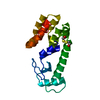
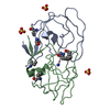
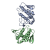
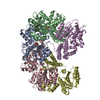
PDB-6tuh:
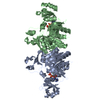
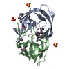
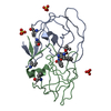
PH domain of Bruton's tyrosine kinase bound to compound 1
Method: X-RAY DIFFRACTION / Resolution: 2.25 Å

PDB-7i90:
Crystal Structure of 7 bound to the PH domain of Btk
Method: X-RAY DIFFRACTION / Resolution: 1.993 Å

PDB-7i91:
Crystal Structure of 13 bound to the PH domain of Btk
Method: X-RAY DIFFRACTION / Resolution: 1.93 Å

PDB-7i92:
Crystal Structure of 9 bound to the PH domain of Btk
Method: X-RAY DIFFRACTION / Resolution: 2.633 Å

PDB-7i93:
Crystal Structure of 27 bound to the PH domain of Btk
Method: X-RAY DIFFRACTION / Resolution: 1.91 Å

PDB-7i94:
Crystal Structure of 33 bound to the PH domain of Btk
Method: X-RAY DIFFRACTION / Resolution: 1.828 Å

PDB-7i95:
Crystal Structure of 25 bound to the PH domain of Btk
Method: X-RAY DIFFRACTION / Resolution: 1.991 Å

PDB-7i96:
Crystal Structure of 4 bound to the PH domain of Btk
Method: X-RAY DIFFRACTION / Resolution: 2.52 Å

PDB-7i97:
Crystal Structure of 8 bound to the PH domain of Btk
Method: X-RAY DIFFRACTION / Resolution: 1.692 Å

PDB-7i98:
Crystal Structure of 10 bound to the PH domain of Btk
Method: X-RAY DIFFRACTION / Resolution: 2.07 Å

PDB-7i99:
Crystal Structure of 22 bound to the PH domain of Btk
Method: X-RAY DIFFRACTION / Resolution: 1.97 Å

PDB-7i9a:
Crystal Structure of 24 bound to the PH domain of Btk
Method: X-RAY DIFFRACTION / Resolution: 2.11 Å

PDB-7i9b:
Crystal Structure of 32 bound to the PH domain of Btk
Method: X-RAY DIFFRACTION / Resolution: 2.769 Å

PDB-7i9c:
Crystal Structure of 30 bound to the PH domain of Btk
Method: X-RAY DIFFRACTION / Resolution: 1.939 Å

PDB-7i9d:
Crystal Structure of 14 bound to the PH domain of Btk
Method: X-RAY DIFFRACTION / Resolution: 1.8 Å

PDB-7i9e:
Crystal Structure of 18 bound to the PH domain of Btk
Method: X-RAY DIFFRACTION / Resolution: 1.91 Å

PDB-7i9f:
Crystal Structure of 16 bound to the PH domain of Btk
Method: X-RAY DIFFRACTION / Resolution: 2.04 Å

PDB-7i9g:
Crystal Structure of 28 bound to the PH domain of Btk
Method: X-RAY DIFFRACTION / Resolution: 1.698 Å

PDB-7i9h:
Crystal Structure of 26 bound to the PH domain of Btk
Method: X-RAY DIFFRACTION / Resolution: 2.589 Å

PDB-7i9i:
Crystal Structure of 2 bound to the PH domain of Btk
Method: X-RAY DIFFRACTION / Resolution: 1.83 Å

PDB-9rl9:
Crystal Structure of 24 bound to the PH domain of Btk
Method: X-RAY DIFFRACTION / Resolution: 2.11 Å

PDB-9rmo:
Crystal Structure of 31 bound to the PH domain of Btk
Method: X-RAY DIFFRACTION / Resolution: 1.8 Å

PDB-9rn5:
Crystal Structure of 33 bound to the PH domain of Btk
Method: X-RAY DIFFRACTION / Resolution: 1.83 Å

PDB-9t0t:
Crystal Structure of the correct enantiomer bound to the PH domain of Btk
Method: X-RAY DIFFRACTION / Resolution: 1.73 Å

PDB-9t1v:
Crystal Structure of 17 bound to the PH domain of Btk
Method: X-RAY DIFFRACTION / Resolution: 2.41 Å

PDB-9t21:
Crystal Structure of 29 bound to the ph domain of Btk
Method: X-RAY DIFFRACTION / Resolution: 1.869 Å

PDB-9t23:
Crystal Structure of 5 bound to the PH domain of Btk
Method: X-RAY DIFFRACTION / Resolution: 1.845 Å

PDB-9t3g:
Crystal Structure of 12 bound to the PH domain of Btk
Method: X-RAY DIFFRACTION / Resolution: 1.946 Å

PDB-9t3m:
Crystal Structure of 11 bound to the PH domain of Btk
Method: X-RAY DIFFRACTION / Resolution: 1.8 Å

Chemicals

ChemComp-NXT:
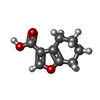
4,5,6,7-tetrahydro-1-benzofuran-3-carboxylic acid

ChemComp-MG:
Unknown entry

ChemComp-ZN:
Unknown entry

ChemComp-HOH:
WATER

PDB-1b6f:
BIRCH POLLEN ALLERGEN BET V 1

PDB-1b6s:
STRUCTURE OF N5-CARBOXYAMINOIMIDAZOLE RIBONUCLEOTIDE SYNTHETASE

PDB-1b6t:
PHOSPHOPANTETHEINE ADENYLYLTRANSFERASE IN COMPLEX WITH 3'-DEPHOSPHO-COA FROM ESCHERICHIA COLI

PDB-1b6u:
CRYSTAL STRUCTURE OF THE HUMAN KILLER CELL INHIBITORY RECEPTOR (KIR2DL3) SPECIFIC FOR HLA-CW3 RELATED ALLELES

PDB-1b6x:
3,N4-ETHENO-2'-DEOXYCYTIDINE OPPOSITE GUANINE IN AN 11-MER DUPLEX, SOLUTION STRUCTURE FROM NMR AND MOLECULAR DYNAMICS, 4 STRUCTURES

PDB-1b6v:
CRYSTAL STRUCTURE OF A HYBRID BETWEEN RIBONUCLEASE A AND BOVINE SEMINAL RIBONUCLEASE

PDB-1b6p:
HIV-1 PROTEASE COMPLEXED WITH MACROCYCLIC PEPTIDOMIMETIC INHIBITOR 7

PDB-1b6q:
ALANINE 31 PROLINE MUTANT OF ROP PROTEIN

PDB-1b6w:
CRYSTAL STRUCTURE OF THE SELENOMETHIONINE VARIANT OF HISTONE HMFB FROM METHANOTHERMUS FERVIDUS

PDB-1b6r:
N5-CARBOXYAMINOIMIDAZOLE RIBONUCLEOTIDE SYNTHETASE FROM E. COLI

PDB-1b6k:
HIV-1 PROTEASE COMPLEXED WITH MACROCYCLIC PEPTIDOMIMETIC INHIBITOR 5

PDB-1b6l:
HIV-1 PROTEASE COMPLEXED WITH MACROCYCLIC PEPTIDOMIMETIC INHIBITOR 4

PDB-1b6m:
HIV-1 PROTEASE COMPLEXED WITH MACROCYCLIC PEPTIDOMIMETIC INHIBITOR 6

ChemComp-CL:
Unknown entry

PDB-1b6g:
HALOALKANE DEHALOGENASE AT PH 5.0 CONTAINING CHLORIDE

PDB-1b6h:
OLIGO-PEPTIDE BINDING PROTEIN COMPLEXED WITH LYSYL-NORVALYL-LYSINE

PDB-1b6i:
T4 LYSOZYME MUTANT WITH CYS 54 REPLACED BY THR, CYS 97 REPLACED BY ALA, THR 21 REPLACED BY CYS AND LYS 124 REPLACED BY CYS (C54T,C97A,T21C,K124C)

PDB-1b6j:
HIV-1 PROTEASE COMPLEXED WITH MACROCYCLIC PEPTIDOMIMETIC INHIBITOR 1

PDB-1jia:
STRUCTURE OF A BASIC PHOSPHOLIPASE A2 FROM AGKISTRODON HALYS PALLAS AT 2.13A RESOLUTION

PDB-1jr3:
Crystal Structure of the Processivity Clamp Loader Gamma Complex of E. coli DNA Polymerase III

PDB-1js1:
Crystal Structure of a new transcarbamylase from the anaerobic bacterium Bacteroides fragilis at 2.0 A resolution

PDB-1js0:
Crystal Structure of 3D Domain-swapped RNase A Minor Trimer

PDB-1js4:
ENDO/EXOCELLULASE:CELLOBIOSE FROM THERMOMONOSPORA

PDB-1js7:
Solution Structure of dAAUAA DNA Bulge

PDB-1js6:
Crystal Structure of DOPA decarboxylase

PDB-1jta:
Crystal Structure of Pectate Lyase A (C2 form)

PDB-1jtb:
LIPID TRANSFER PROTEIN COMPLEXED WITH PALMITOYL COENZYME A, NMR, 16 STRUCTURES

Source
  • homo sapiens (human)
KeywordsTRANSFERASE / BTK / Covalent fragments / surface entrophy reduction / crystal engineering / HYDROLASE / Fragment based drug discovery / ph domain

+
About Yorodumi Papers

-
News

-
Feb 9, 2022. New format data for meta-information of EMDB entries

New format data for meta-information of EMDB entries

  • Version 3 of the EMDB header file is now the official format.
  • The previous official version 1.9 will be removed from the archive.

Related info.:EMDB header

External links:wwPDB to switch to version 3 of the EMDB data model

-
Aug 12, 2020. Covid-19 info

Covid-19 info

URL: https://pdbj.org/emnavi/covid19.php

New page: Covid-19 featured information page in EM Navigator.

Related info.:Covid-19 info / Mar 5, 2020. Novel coronavirus structure data

+
Mar 5, 2020. Novel coronavirus structure data

Novel coronavirus structure data

Related info.:Yorodumi Speices / Aug 12, 2020. Covid-19 info

External links:COVID-19 featured content - PDBj / Molecule of the Month (242):Coronavirus Proteases

+
Jan 31, 2019. EMDB accession codes are about to change! (news from PDBe EMDB page)

EMDB accession codes are about to change! (news from PDBe EMDB page)

  • The allocation of 4 digits for EMDB accession codes will soon come to an end. Whilst these codes will remain in use, new EMDB accession codes will include an additional digit and will expand incrementally as the available range of codes is exhausted. The current 4-digit format prefixed with “EMD-” (i.e. EMD-XXXX) will advance to a 5-digit format (i.e. EMD-XXXXX), and so on. It is currently estimated that the 4-digit codes will be depleted around Spring 2019, at which point the 5-digit format will come into force.
  • The EM Navigator/Yorodumi systems omit the EMD- prefix.

Related info.:Q: What is EMD? / ID/Accession-code notation in Yorodumi/EM Navigator

External links:EMDB Accession Codes are Changing Soon! / Contact to PDBj

+
Jul 12, 2017. Major update of PDB

Major update of PDB

  • wwPDB released updated PDB data conforming to the new PDBx/mmCIF dictionary.
  • This is a major update changing the version number from 4 to 5, and with Remediation, in which all the entries are updated.
  • In this update, many items about electron microscopy experimental information are reorganized (e.g. em_software).
  • Now, EM Navigator and Yorodumi are based on the updated data.

External links:wwPDB Remediation / Enriched Model Files Conforming to OneDep Data Standards Now Available in the PDB FTP Archive

-
Yorodumi Papers

Database of articles cited by EMDB/PDB/SASBDB data

  • Database of articles cited by EMDB, PDB, and SASBDB entries
  • Using PubMed data

Related info.:EMDB / PDB / SASBDB / Yorodumi / EMN Papers / Changes in new EM Navigator and Yorodumi

Read more