 Movie
Movie Controller

[English] 日本語
 Yorodumi
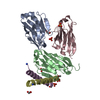
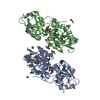
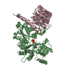
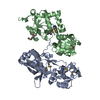
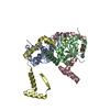
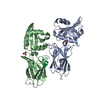
- PDB-1b6u: CRYSTAL STRUCTURE OF THE HUMAN KILLER CELL INHIBITORY RECEPTOR (K... -
+ Open data

- Basic information
| Entry | Database: PDB / ID: 1b6u | ||||||
|---|---|---|---|---|---|---|---|
| Title | CRYSTAL STRUCTURE OF THE HUMAN KILLER CELL INHIBITORY RECEPTOR (KIR2DL3) SPECIFIC FOR HLA-CW3 RELATED ALLELES | ||||||
 Components Components | P58 KILLER CELL INHIBITORY RECEPTOR | ||||||
 Keywords Keywords | KILLER CELL INHIBITORY RECEPTOR / NATURAL KILLER CELL / HLA / MAJOR HISTOCOMPATIBILITY COMPLEX CLASS I (MHC CLASS I) / CELL SURFACE RECEPTOR / IMMUNOGLOBULIN SUPERFAMILY | ||||||
| Function / homology |  Function and homology information Function and homology informationimmune response-regulating signaling pathway / antigen binding / Immunoregulatory interactions between a Lymphoid and a non-Lymphoid cell / signaling receptor activity / immune response / membrane / identical protein binding / plasma membrane Similarity search - Function | ||||||
| Biological species |  Homo sapiens (human) Homo sapiens (human) | ||||||
| Method |  X-RAY DIFFRACTION / SYNCHROTRON / MOLECULAR REPLACEMENT / Resolution: 3 Å X-RAY DIFFRACTION / SYNCHROTRON / MOLECULAR REPLACEMENT / Resolution: 3 Å | ||||||
 Authors Authors | Maenaka, K. / Juji, T. / Stuart, D.I. / Jones, E.Y. | ||||||
 Citation Citation | Journal: Structure Fold.Des. / Year: 1999 Title: Crystal structure of the human p58 killer cell inhibitory receptor (KIR2DL3) specific for HLA-Cw3-related MHC class I. Authors: Maenaka, K. / Juji, T. / Stuart, D.I. / Jones, E.Y. | ||||||
| History |
|
- Structure visualization
| Structure viewer | Molecule: MolmilJmol/JSmol |
|---|
- Downloads & links
-Download
| PDBx/mmCIF format | 1b6u.cif.gz | 50.3 KB | Display | PDBx/mmCIF format |
|---|---|---|---|---|
| PDB format | pdb1b6u.ent.gz | 36.6 KB | Display | PDB format |
| PDBx/mmJSON format | 1b6u.json.gz | Tree view | PDBx/mmJSON format | |
| Others |  Other downloads Other downloads |
-Validation report
| Arichive directory | https://data.pdbj.org/pub/pdb/validation_reports/b6/1b6uftp://data.pdbj.org/pub/pdb/validation_reports/b6/1b6u | HTTPS FTP |
|---|
-Related structure data
| Similar structure data |
|---|


-Links
 PDBj
PDBj


- Assembly
| Deposited unit | 
| ||||||||
|---|---|---|---|---|---|---|---|---|---|
| 1 | 
| ||||||||
| Unit cell |
|
-Components
| #1: Protein | Mass: 28189.141 Da / Num. of mol.: 1 / Fragment: EXTRACELLULAR REGION Source method: isolated from a genetically manipulated source Source: (gene. exp.) Homo sapiens (human) / Description: CLONING BY PCR / Cell: NATURAL KILLER CELL / Cell line: BL21 / Cellular location: CELL SURFACE / Gene: NKAT2 / Plasmid: PKMATHNK2Cellular location (production host): MEDIA AND PERIPLASMIC SPACE Gene (production host): NKAT2 / Production host:  |
|---|---|
| Has protein modification | Y |
-Experimental details
-Experiment
| Experiment | Method: X-RAY DIFFRACTION / Number of used crystals: 1 |
|---|
- Sample preparation
| Crystal | Density Matthews: 3.05 Å3/Da / Density % sol: 60 % / Description: DATA WERE COLLECTED USING WEISSENBERG METHOD. | |||||||||||||||||||||||||||||||||||
|---|---|---|---|---|---|---|---|---|---|---|---|---|---|---|---|---|---|---|---|---|---|---|---|---|---|---|---|---|---|---|---|---|---|---|---|---|
| Crystal grow | pH: 7.5 / Details: pH 7.5 | |||||||||||||||||||||||||||||||||||
| Crystal grow | *PLUS Temperature: 21 ℃ / Method: vapor diffusion, sitting dropDetails: protein solution is mixed in a 1:1 ratio with well solution | |||||||||||||||||||||||||||||||||||
| Components of the solutions | *PLUS
|
-Data collection
| Diffraction | Mean temperature: 288 K |
|---|---|
| Diffraction source | Source: SYNCHROTRON / Site: Photon Factory  / Beamline: BL-6A / Wavelength: 1 / Wavelength: 1 Å / Beamline: BL-6A / Wavelength: 1 / Wavelength: 1 Å |
| Detector | Type: FUJI / Detector: IMAGE PLATE / Date: Oct 1, 1996 / Details: MIRRORS |
| Radiation | Protocol: SINGLE WAVELENGTH / Monochromatic (M) / Laue (L): M / Scattering type: x-ray |
| Radiation wavelength | Wavelength: 1 Å / Relative weight: 1 |
| Reflection | Resolution: 3→20 Å / Num. obs: 6281 / % possible obs: 84.9 % / Observed criterion σ(I): 0 / Redundancy: 2.9 % / Rmerge(I) obs: 0.195 / Net I/σ(I): 4.7 |
| Reflection shell | Resolution: 3→3.11 Å / Rmerge(I) obs: 0.49 / Mean I/σ(I) obs: 1.5 / % possible all: 87.6 |
| Reflection | *PLUS Num. measured all: 18460 |
| Reflection shell | *PLUS % possible obs: 87.6 % |
- Processing
| Software |
| ||||||||||||||||||||||||||||||||||||||||||||||||||||||||||||
|---|---|---|---|---|---|---|---|---|---|---|---|---|---|---|---|---|---|---|---|---|---|---|---|---|---|---|---|---|---|---|---|---|---|---|---|---|---|---|---|---|---|---|---|---|---|---|---|---|---|---|---|---|---|---|---|---|---|---|---|---|---|
| Refinement | Method to determine structure: MOLECULAR REPLACEMENT / Resolution: 3→15 Å / Cross valid method: THROUGHOUT / σ(F): 0 / Details: REFINED BY X-PLOR, REFMAC AND CN
| ||||||||||||||||||||||||||||||||||||||||||||||||||||||||||||
| Solvent computation | Solvent model: FLAT MODEL / Bsol: 20 Å2 / ksol: 0.25 e/Å3 | ||||||||||||||||||||||||||||||||||||||||||||||||||||||||||||
| Displacement parameters | Biso mean: 40.8 Å2
| ||||||||||||||||||||||||||||||||||||||||||||||||||||||||||||
| Refinement step | Cycle: LAST / Resolution: 3→15 Å
| ||||||||||||||||||||||||||||||||||||||||||||||||||||||||||||
| Refine LS restraints |
| ||||||||||||||||||||||||||||||||||||||||||||||||||||||||||||
| Software | *PLUS Name: CNS / Version: 0.5 / Classification: refinement | ||||||||||||||||||||||||||||||||||||||||||||||||||||||||||||
| Refinement | *PLUS | ||||||||||||||||||||||||||||||||||||||||||||||||||||||||||||
| Solvent computation | *PLUS | ||||||||||||||||||||||||||||||||||||||||||||||||||||||||||||
| Displacement parameters | *PLUS |
