 Movie
Movie Controller
 Structure viewers
 About EMN search


-Search query
-Search result
Showing 1 - 50 of 165 items for (author: smith & sp)

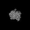
EMDB-71766: 
Cryo-EM structure of J601-1B2 Fab in complex with HIV-1 BG505 DS-SOSIP Env trimer
Method: single particle / : Wang S, Zhou T, Kwong PD

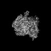
EMDB-71767:
Cryo-EM structure of J601-A6 Fab in complex with HIV-1 BG505 DS-SOSIP Env trimer
Method: single particle / : Wang S, Zhou T, Kwong PD, Morano NC, Shapiro L

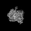
EMDB-71772:
Cryo-EM structure of K001-A1 Fab in complex with HIV-1 459C-OPT RnS DS-SOSIP Env trimer
Method: single particle / : Wang S, Zhou T, Kwong PD, Morano NC, Shapiro L

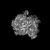
EMDB-71781:
Cryo-EM structure of HIV-1 459C-WT DS-SOSIP RnS Env trimer
Method: single particle / : Wang S, Zhou T, Kwong PD, Morano NC, Shapiro L

EMDB-71782:
Cryo-EM structure of HIV-1 459C-ALT DS-SOSIP RnS Env trimer
Method: single particle / : Wang S, Zhou T, Kwong PD, Morano NC, Shapiro L

PDB-9pni:
Cryo-EM structure of J601-1B2 Fab in complex with HIV-1 BG505 DS-SOSIP Env trimer
Method: single particle / : Wang S, Zhou T, Kwong PD

PDB-9pnn:
Cryo-EM structure of J601-A6 Fab in complex with HIV-1 BG505 DS-SOSIP Env trimer
Method: single particle / : Wang S, Zhou T, Kwong PD, Morano NC, Shapiro L

PDB-9pnu:
Cryo-EM structure of K001-A1 Fab in complex with HIV-1 459C-OPT RnS DS-SOSIP Env trimer
Method: single particle / : Wang S, Zhou T, Kwong PD, Morano NC, Shapiro L

PDB-9pq2:
Cryo-EM structure of HIV-1 459C-WT DS-SOSIP RnS Env trimer
Method: single particle / : Wang S, Zhou T, Kwong PD, Morano NC, Shapiro L

PDB-9pq3:
Cryo-EM structure of HIV-1 459C-ALT DS-SOSIP RnS Env trimer
Method: single particle / : Wang S, Zhou T, Kwong PD, Morano NC, Shapiro L

EMDB-46824:
Polyclonal immune complex of human subject 321-2006 Fab binding H1 HA
Method: single particle / : Han J, Rodriguez AJ, Ferguson JA, Ward AB

EMDB-46825:
Polyclonal immune complex of human subject 321-2009 Fab binding H1 HA
Method: single particle / : Han J, Rodriguez AJ, Ferguson JA, Ward AB

EMDB-46827:
Polyclonal immune complex of human subject 321-2012 Fab binding H1 HA
Method: single particle / : Han J, Rodriguez AJ, Ferguson JA, Ward AB

EMDB-46829:
Polyclonal immune complex of Fab from Cynomolgus Macaque 6974 at week 12 binding H1 HA
Method: single particle / : Han J, Rodriguez AJ, Ferguson JA, Ward AB

EMDB-46830:
Polyclonal immune complex of Fab from Rhesus Macaque BB798E at week 12 binding H1 HA
Method: single particle / : Han J, Rodriguez AJ, Ferguson JA, Ward AB

EMDB-46831:
Polyclonal immune complex of Fab from Cynomolgus Macaque T009 at week 12 binding H1 HA
Method: single particle / : Han J, Rodriguez AJ, Ferguson JA, Ward AB

EMDB-46832:
Polyclonal immune complex of Fab from Cynomolgus Macaque R996 at week 12 binding H1 HA
Method: single particle / : Han J, Rodriguez AJ, Ferguson JA, Ward AB

EMDB-45636:
CryoEM structure of NC99 hemagglutinin trimer in complex with Fab BB798E 3-C07
Method: single particle / : Li N, Tsybovsky Y, Sangesland M, Kanekiyo M

EMDB-45637:
CryoEM structure of NC99 hemagglutinin trimer in complex with Fab T009 3-E04
Method: single particle / : Li N, Tsybovsky Y, Sangesland M, Kanekiyo M

PDB-9cjy:
CryoEM structure of NC99 hemagglutinin trimer in complex with Fab BB798E 3-C07
Method: single particle / : Li N, Tsybovsky Y, Sangesland M, Kanekiyo M

PDB-9cjz:
CryoEM structure of NC99 hemagglutinin trimer in complex with Fab T009 3-E04
Method: single particle / : Li N, Tsybovsky Y, Sangesland M, Kanekiyo M
EMDB-50675:
Escherichia coli 70S ribosome in situ structure
Method: subtomogram averaging / : Watson H, Berger C, Grange M
EMDB-50676:
E. coli 70S ribosome in situ structure for xenon damage layer determination: 5 - 10 nm
Method: subtomogram averaging / : Watson H, Berger C, Grange M
EMDB-50678:
E. coli 70S ribosome in situ structure for xenon damage layer determination: 10 - 15 nm
Method: subtomogram averaging / : Watson H, Berger C, Grange M
EMDB-50679:
E. coli 70S ribosome in situ structure for xenon damage layer determination: 15 - 20 nm
Method: subtomogram averaging / : Watson H, Berger C, Grange M

EMDB-50680:
E. coli 70S ribosome in situ structure for xenon damage layer determination: 20 - 25 nm
Method: subtomogram averaging / : Watson H, Berger C, Grange M

EMDB-50681:
E. coli 70S ribosome in situ structure for xenon damage layer determination: 25 - 30 nm
Method: subtomogram averaging / : Watson H, Berger C, Grange M
EMDB-50682:
E. coli 70S ribosome in situ structure for xenon damage layer determination: 30 - 35 nm
Method: subtomogram averaging / : Watson H, Berger C, Grange M
EMDB-50683:
E. coli 70S ribosome in situ structure for xenon damage layer determination: 35 - 40 nm
Method: subtomogram averaging / : Watson H, Berger C, Grange M
EMDB-50684:
E. coli 70S ribosome in situ structure for xenon damage layer determination: 40 - 45 nm
Method: subtomogram averaging / : Watson H, Berger C, Grange M
EMDB-50685:
E. coli 70S ribosome in situ structure for xenon damage layer determination: 45 - 50 nm
Method: subtomogram averaging / : Watson H, Berger C, Grange M
EMDB-50686:
E. coli 70S ribosome in situ structure for xenon damage layer determination: 50 - 55 nm
Method: subtomogram averaging / : Watson H, Berger C, Grange M

EMDB-50687:
E. coli 70S ribosome in situ structure for xenon damage layer determination: 55 - 60 nm
Method: subtomogram averaging / : Watson H, Berger C, Grange M
EMDB-50688:
E. coli 70S ribosome in situ structure for xenon damage layer determination: >10nm matched control for 5 - 10 nm
Method: subtomogram averaging / : Watson H, Berger C, Grange M

EMDB-50689:
E. coli 70S ribosome in situ structure for xenon damage layer determination: >15 nm matched control for 10 - 15 nm
Method: subtomogram averaging / : Watson H, Berger C, Grange M

EMDB-50690:
E. coli 70S ribosome in situ structure for xenon damage layer determination: >20 nm matched control for 15 - 20 nm
Method: subtomogram averaging / : Watson H, Berger C, Grange M

EMDB-50691:
E. coli 70S ribosome in situ structure for xenon damage layer determination: >25 nm matched control for 20 - 25 nm
Method: subtomogram averaging / : Watson H, Berger C, Grange M

EMDB-50692:
E. coli 70S ribosome in situ structure for xenon damage layer determination: >30 nm matched control for 25 - 30 nm
Method: subtomogram averaging / : Watson H, Berger C, Grange M

EMDB-50693:
E. coli 70S ribosome in situ structure for xenon damage layer determination: >35 nm matched control for 30 - 35 nm
Method: subtomogram averaging / : Watson H, Berger C, Grange M

EMDB-50694:
E. coli 70S ribosome in situ structure for xenon damage layer determination: >40 nm matched control for 35 - 40 nm
Method: subtomogram averaging / : Watson H, Berger C, Grange M

EMDB-50695:
E. coli 70S ribosome in situ structure for xenon damage layer determination: >45 nm matched control for 40 - 45 nm
Method: subtomogram averaging / : Watson H, Berger C, Grange M

EMDB-50696:
E. coli 70S ribosome in situ structure for xenon damage layer determination: >50 nm matched control for 45 - 50 nm
Method: subtomogram averaging / : Watson H, Berger C, Grange M

EMDB-50697:
E. coli 70S ribosome in situ structure for xenon damage layer determination: >55 nm matched control for 50 - 55 nm
Method: subtomogram averaging / : Watson H, Berger C, Grange M

EMDB-50698:
E. coli 70S ribosome in situ structure for xenon damage layer determination: >60 nm matched control for 55 - 60 nm
Method: subtomogram averaging / : Watson H, Berger C, Grange M

EMDB-50699:
E. coli 70S ribosome in situ structure for lamellae backside damage analysis: 0 - 1 micron from lamellae backside
Method: subtomogram averaging / : Watson H, Berger C, Grange M

EMDB-50700:
E. coli 70S ribosome in situ structure for lamellae backside damage analysis: 1 - 2 microns from lamellae backside
Method: subtomogram averaging / : Watson H, Berger C, Grange M

EMDB-50701:
E. coli 70S ribosome in situ structure for lamellae backside damage analysis: 2 - 3 microns from lamellae backside
Method: subtomogram averaging / : Watson H, Berger C, Grange M

EMDB-50702:
E. coli 70S ribosome in situ structure for lamellae backside damage analysis: 3 - 4 microns from lamellae backside
Method: subtomogram averaging / : Watson H, Berger C, Grange M

EMDB-50703:
E. coli 70S ribosome in situ structure for lamellae backside damage analysis: 4 - 5 microns from lamellae backside
Method: subtomogram averaging / : Watson H, Berger C, Grange M

EMDB-50704:
E. coli 70S ribosome in situ structure for xenon damage layer determination: 10,000 particles (no depth constraint)
Method: subtomogram averaging / : Watson H, Berger C, Grange M
Pages:
 wwPDB to switch to version 3 of the EMDB data model
