 Movie
Movie Controller
 Structure viewers
 About EMN search


-Search query
-Search result
Showing all 32 items for (author: rutten & t)

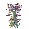
EMDB-71766: 
Cryo-EM structure of J601-1B2 Fab in complex with HIV-1 BG505 DS-SOSIP Env trimer
Method: single particle / : Wang S, Zhou T, Kwong PD

EMDB-71767:
Cryo-EM structure of J601-A6 Fab in complex with HIV-1 BG505 DS-SOSIP Env trimer
Method: single particle / : Wang S, Zhou T, Kwong PD, Morano NC, Shapiro L

EMDB-71772:
Cryo-EM structure of K001-A1 Fab in complex with HIV-1 459C-OPT RnS DS-SOSIP Env trimer
Method: single particle / : Wang S, Zhou T, Kwong PD, Morano NC, Shapiro L

EMDB-71781:
Cryo-EM structure of HIV-1 459C-WT DS-SOSIP RnS Env trimer
Method: single particle / : Wang S, Zhou T, Kwong PD, Morano NC, Shapiro L

EMDB-71782:
Cryo-EM structure of HIV-1 459C-ALT DS-SOSIP RnS Env trimer
Method: single particle / : Wang S, Zhou T, Kwong PD, Morano NC, Shapiro L

PDB-9pni:
Cryo-EM structure of J601-1B2 Fab in complex with HIV-1 BG505 DS-SOSIP Env trimer
Method: single particle / : Wang S, Zhou T, Kwong PD

PDB-9pnn:
Cryo-EM structure of J601-A6 Fab in complex with HIV-1 BG505 DS-SOSIP Env trimer
Method: single particle / : Wang S, Zhou T, Kwong PD, Morano NC, Shapiro L

PDB-9pnu:
Cryo-EM structure of K001-A1 Fab in complex with HIV-1 459C-OPT RnS DS-SOSIP Env trimer
Method: single particle / : Wang S, Zhou T, Kwong PD, Morano NC, Shapiro L

PDB-9pq2:
Cryo-EM structure of HIV-1 459C-WT DS-SOSIP RnS Env trimer
Method: single particle / : Wang S, Zhou T, Kwong PD, Morano NC, Shapiro L

PDB-9pq3:
Cryo-EM structure of HIV-1 459C-ALT DS-SOSIP RnS Env trimer
Method: single particle / : Wang S, Zhou T, Kwong PD, Morano NC, Shapiro L
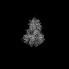
EMDB-42060:
Cryo-EM structure of prefusion-stabilized influenza B hemagglutinin
Method: single particle / : Juraszek J, Milder FJ, Yu X, Blokland S, Overveld D, Tamara S, Bakkers MJG, Rutten L, Sharma S, Langedijk JPM
EMDB-43273:
Prefusion-stabilized trimer of FluB-HA under low pH conditions
Method: single particle / : Juraszek J, Milder FJ, Yu X, Blokland S, Overveld D, Tamara S, Bakkers MJG, Rutten L, Sharma S, Langedijk JPM
PDB-8uad:
Cryo-EM structure of prefusion-stabilized influenza B hemagglutinin
Method: single particle / : Juraszek J, Milder FJ, Yu X, Blokland S, Overveld D, Tamara S, Bakkers MJG, Rutten L, Sharma S, Langedijk JPM
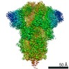
EMDB-29454:
Structure of Covid Spike variant deltaN135 in fully closed form
Method: single particle / : Yu X, Juraszek J, Rutten L, Bakkers MJG, Blokland S, Van den Broek NJF, Verwilligen AYW, Abeywickrema P, Vingerhoets J, Neefs J, Bakhash SAM, Roychoudhury P, Greninger A, Sharma S, Langedijk JPM
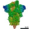
EMDB-29455:
Structure of Covid Spike variant deltaN135 with one erect RBD
Method: single particle / : Yu X, Juraszek J, Rutten L, Bakkers MJG, Blokland S, Van den Broek NJF, Verwilligen AYW, Abeywickrema P, Vingerhoets J, Neefs J, Bakhash SAM, Roychoudhury P, Greninger A, Sharma S, Langedijk JPM

EMDB-29456:
Structure of Covid Spike variant deltaN25 with one erect RBD
Method: single particle / : Yu X, Juraszek J, Rutten L, Bakkers MJG, Blokland S, Van den Broek NJF, Verwilligen AYW, Abeywickrema P, Vingerhoets J, Neefs J, Bakhash SAM, Roychoudhury P, Greninger A, Sharma S, Langedijk JPM
PDB-8fu7:
Structure of Covid Spike variant deltaN135 in fully closed form
Method: single particle / : Yu X, Juraszek J, Rutten L, Bakkers MJG, Blokland S, Van den Broek NJF, Verwilligen AYW, Abeywickrema P, Vingerhoets J, Neefs J, Bakhash SAM, Roychoudhury P, Greninger A, Sharma S, Langedijk JPM
PDB-8fu8:
Structure of Covid Spike variant deltaN135 with one erect RBD
Method: single particle / : Yu X, Juraszek J, Rutten L, Bakkers MJG, Blokland S, Van den Broek NJF, Verwilligen AYW, Abeywickrema P, Vingerhoets J, Neefs J, Bakhash SAM, Roychoudhury P, Greninger A, Sharma S, Langedijk JPM
PDB-8fu9:
Structure of Covid Spike variant deltaN25 with one erect RBD
Method: single particle / : Yu X, Juraszek J, Rutten L, Bakkers MJG, Blokland S, Van den Broek NJF, Verwilligen AYW, Abeywickrema P, Vingerhoets J, Neefs J, Bakhash SAM, Roychoudhury P, Greninger A, Sharma S, Langedijk JPM

EMDB-23480:
Cryo-EM structure of llama J3 VHH antibody in complex with HIV-1 Env BG505 DS-SOSIP.664
Method: single particle / : Gorman J, Kwong PD

PDB-7lpn:
Cryo-EM structure of llama J3 VHH antibody in complex with HIV-1 Env BG505 DS-SOSIP.664
Method: single particle / : Gorman J, Kwong PD
EMDB-11639:
Cryo-EM structure of a prefusion stabilized SARS-CoV-2 Spike (D614N, R682S, R685G, A892P, A942P and V987P)(S-closed trimer)
Method: single particle / : Renault LLR, Rutten L, Juraszek J, Langedijk JPM
EMDB-11719:
Cryo-EM structure of a prefusion stabilized SARS-CoV-2 Spike (D614N, R682S, R685G, A892P, A942P and V987P)(One up trimer)
Method: single particle / : Renault LLR, Rutten L, Juraszek J, Langedijk JPM

PDB-7a4n:
Cryo-EM structure of a prefusion stabilized SARS-CoV-2 Spike (D614N, R682S, R685G, A892P, A942P and V987P)(S-closed trimer)
Method: single particle / : Rutten L, Renault LLR, Juraszek J, Langedijk JPM

PDB-7ad1:
Cryo-EM structure of a prefusion stabilized SARS-CoV-2 Spike (D614N, R682S, R685G, A892P, A942P and V987P)(One up trimer)
Method: single particle / : Rutten L, Renault LLR, Juraszek J, Langedijk JPM
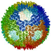
EMDB-1015:
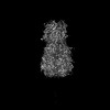
Cryo-electron microscopy reveals the functional organization of an enveloped virus, Semliki Forest virus.
Method: single particle / : Mancini EJ, Clarke M, Gowen B, Rutten T, Fuller SD
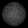
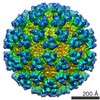
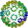
EMDB-1013:
Combined EM/X-ray imaging yields a quasi-atomic model of the adenovirus-related bacteriophage PRD1 and shows key capsid and membrane interactions.
Method: single particle / : San Martin C, Burnett RM, de Haas F, Heinkel R, Rutten T, Fuller SD, Butcher SJ, Bamford DH
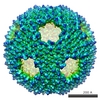
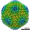
EMDB-1014:
Combined EM/X-ray imaging yields a quasi-atomic model of the adenovirus-related bacteriophage PRD1 and shows key capsid and membrane interactions.
Method: single particle / : San Martin C, Burnett RM, de Haas F, Heinkel R, Rutten T, Fuller SD, Butcher SJ, Bamford DH
PDB-1hb5:
quasi-atomic resolution model of bacteriophage PRD1 P3-shell, obtained by combined cryo-EM and X-ray crystallography.
Method: single particle / : San Martin C, Burnett RM, De Haas F, Heinkel R, Rutten T, Fuller SD, Butcher SJ, Bamford DH
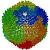
PDB-1hb7:
quasi-atomic resolution model of bacteriophage PRD1 sus1 mutant, obtained by combined cryo-EM and X-ray crystallography.
Method: single particle / : San Martin C, Burnett RM, De Haas F, Heinkel R, Rutten T, Fuller SD, Butcher SJ, Bamford DH
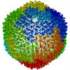
PDB-1hb9:
quasi-atomic resolution model of bacteriophage PRD1 wild type virion, obtained by combined cryo-EM and X-ray crystallography.
Method: single particle / : San Martin C, Burnett RM, De Haas F, Heinkel R, Rutten T, Fuller SD, Butcher SJ, Bamford DH
 wwPDB to switch to version 3 of the EMDB data model
