 Movie
Movie Controller

[English] 日本語
 Yorodumi
- EMDB-42060: Cryo-EM structure of prefusion-stabilized influenza B hemagglutinin -
+ Open data

- Basic information
| Entry |  | |||||||||
|---|---|---|---|---|---|---|---|---|---|---|
| Title | Cryo-EM structure of prefusion-stabilized influenza B hemagglutinin | |||||||||
 Map data Map data | ||||||||||
 Sample Sample |
| |||||||||
 Keywords Keywords | Cryo-EM / FluB / HA / VIRAL PROTEIN | |||||||||
| Function / homology |  Function and homology information Function and homology informationviral budding from plasma membrane / host cell surface receptor binding / endocytosis involved in viral entry into host cell / fusion of virus membrane with host plasma membrane / fusion of virus membrane with host endosome membrane / viral envelope / virion attachment to host cell / host cell plasma membrane / virion membrane / membrane Similarity search - Function | |||||||||
| Biological species |  Influenza B virus Influenza B virus | |||||||||
| Method | single particle reconstruction / cryo EM / Resolution: 2.77 Å | |||||||||
 Authors Authors | Juraszek J / Milder FJ / Yu X / Blokland S / Overveld D / Tamara S / Bakkers MJG / Rutten L / Sharma S / Langedijk JPM | |||||||||
| Funding support | 1 items
| |||||||||
 Citation Citation | Journal: PNAS Nexus / Year: 2024 Title: Engineering a cleaved, prefusion-stabilized influenza B virus hemagglutinin by identification and locking of all six pH switches. Authors: Jarek Juraszek / Fin J Milder / Xiaodi Yu / Sven Blokland / Daan van Overveld / Pravien Abeywickrema / Sem Tamara / Sujata Sharma / Lucy Rutten / Mark J G Bakkers / Johannes P M Langedijk /   Abstract: Vaccine components based on viral fusion proteins require high stability of the native prefusion conformation for optimal potency and manufacturability. In the case of influenza B virus hemagglutinin ...Vaccine components based on viral fusion proteins require high stability of the native prefusion conformation for optimal potency and manufacturability. In the case of influenza B virus hemagglutinin (HA), the stem's conformation relies on efficient cleavage. In this study, we identified six pH-sensitive regions distributed across the entire ectodomain where protonated histidines assume either a repulsive or an attractive role. Substitutions in these areas enhanced the protein's expression, quality, and stability in its prefusion trimeric state. Importantly, this stabilization enabled the production of a cleavable HA0, which is further processed into HA1 and HA2 by furin during exocytic pathway passage, thereby facilitating correct folding, increased stability, and screening for additional stabilizing substitutions in the core of the metastable fusion domain. Cryo-EM analysis at neutral and low pH revealed a previously unnoticed pH switch involving the C-terminal residues of the natively cleaved HA1. This switch keeps the fusion peptide in a clamped state at neutral pH, averting premature conformational shift. Our findings shed light on new strategies for possible improvements of recombinant or genetic-based influenza B vaccines. | |||||||||
| History |
|
- Structure visualization
| Supplemental images |
|---|

- Downloads & links
-EMDB archive
| Map data | emd_42060.map.gz | 79 MB | EMDB map data format | |
|---|---|---|---|---|
| Header (meta data) | emd-42060-v30.xmlemd-42060.xml | 20.4 KB 20.4 KB | Display Display | EMDB header |
| Images |  emd_42060.png emd_42060.png | 30.8 KB | ||
| Masks | emd_42060_msk_1.map | 83.7 MB | Mask map | |
| Filedesc metadata | emd-42060.cif.gz | 6.3 KB | ||
| Others | emd_42060_additional_1.map.gzemd_42060_half_map_1.map.gzemd_42060_half_map_2.map.gz | 41.7 MB 77.7 MB 77.7 MB | ||
| Archive directory |  http://ftp.pdbj.org/pub/emdb/structures/EMD-42060ftp://ftp.pdbj.org/pub/emdb/structures/EMD-42060 http://ftp.pdbj.org/pub/emdb/structures/EMD-42060ftp://ftp.pdbj.org/pub/emdb/structures/EMD-42060 | HTTPS FTP |
-Related structure data
| Related structure data | 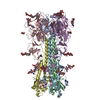 8uadMC M: atomic model generated by this map C: citing same article ( |
|---|---|
| Similar structure data |
-Links
| EMDB pages | EMDB (EBI/PDBe) / EMDataResource |
|---|---|
| Related items in Molecule of the Month |


-Map
| File | Download / File: emd_42060.map.gz / Format: CCP4 / Size: 83.7 MB / Type: IMAGE STORED AS FLOATING POINT NUMBER (4 BYTES) | ||||||||||||||||||||||||||||||||||||
|---|---|---|---|---|---|---|---|---|---|---|---|---|---|---|---|---|---|---|---|---|---|---|---|---|---|---|---|---|---|---|---|---|---|---|---|---|---|
| Projections & slices | Image control
Images are generated by Spider. | ||||||||||||||||||||||||||||||||||||
| Voxel size | X=Y=Z: 0.91 Å | ||||||||||||||||||||||||||||||||||||
| Density |
| ||||||||||||||||||||||||||||||||||||
| Symmetry | Space group: 1 | ||||||||||||||||||||||||||||||||||||
| Details | EMDB XML:
|
 Z (Sec.)
 Y (Row.)
 X (Col.)


-Supplemental data
-Mask #1
| File | emd_42060_msk_1.map | ||||||||||||
|---|---|---|---|---|---|---|---|---|---|---|---|---|---|
| Projections & Slices |
| ||||||||||||
| Density Histograms |


-Additional map: #1
| File | emd_42060_additional_1.map | ||||||||||||
|---|---|---|---|---|---|---|---|---|---|---|---|---|---|
| Projections & Slices |
| ||||||||||||
| Density Histograms |


-Half map: #1
| File | emd_42060_half_map_1.map | ||||||||||||
|---|---|---|---|---|---|---|---|---|---|---|---|---|---|
| Projections & Slices |
| ||||||||||||
| Density Histograms |


-Half map: #2
| File | emd_42060_half_map_2.map | ||||||||||||
|---|---|---|---|---|---|---|---|---|---|---|---|---|---|
| Projections & Slices |
| ||||||||||||
| Density Histograms |


- Sample components
-Entire : prefusion-stabilized influenza B hemagglutinin
| Entire | Name: prefusion-stabilized influenza B hemagglutinin |
|---|---|
| Components |
|
-Supramolecule #1: prefusion-stabilized influenza B hemagglutinin
| Supramolecule | Name: prefusion-stabilized influenza B hemagglutinin / type: complex / ID: 1 / Parent: 0 / Macromolecule list: #1-#2 |
|---|---|
| Source (natural) | Organism: Influenza B virus |
-Macromolecule #1: Hemagglutinin HA1 chain
| Macromolecule | Name: Hemagglutinin HA1 chain / type: protein_or_peptide / ID: 1 / Number of copies: 3 / Enantiomer: LEVO |
|---|---|
| Source (natural) | Organism: Influenza B virus |
| Molecular weight | Theoretical: 38.928676 KDa |
| Recombinant expression | Organism:  Homo sapiens (human) Homo sapiens (human) |
| Sequence | String: MKAIIVLLMV VTSNADRICT GITSSNSPHV VKTATQGEVN VTGVIPLTTT PTKSHFANLK GTETRGKLCP KCLNCTDLDV ALGRPKCTG KIPSARVSIL HEVRPVTSGC FPIMHDRTKI RQLPNLLRGY EHVRLSTHNV INAEGAPGGP YKIGTSGSCP N ITNGNGFF ...String: MKAIIVLLMV VTSNADRICT GITSSNSPHV VKTATQGEVN VTGVIPLTTT PTKSHFANLK GTETRGKLCP KCLNCTDLDV ALGRPKCTG KIPSARVSIL HEVRPVTSGC FPIMHDRTKI RQLPNLLRGY EHVRLSTHNV INAEGAPGGP YKIGTSGSCP N ITNGNGFF ATMAWAVPDK NKTATNPLTI EVPYVCTEGE DQITVWGFHS DNETQMAKLY GDSKPQTFTS SANGVTTHYV SQ IGGFPNQ TEDGGLPQSG RIVVDYMVQK SGKTGTITYQ RGILLPQKVW CASGRSKVIK GSLPLIGEAD CLHEKYGGLN KSK PYYTGE HAKAIGNCPI WVKTPLKLAN GTKYRPPAKL LKER UniProtKB: Hemagglutinin |
-Macromolecule #2: Hemagglutinin HA2 chain
| Macromolecule | Name: Hemagglutinin HA2 chain / type: protein_or_peptide / ID: 2 / Number of copies: 3 / Enantiomer: LEVO |
|---|---|
| Source (natural) | Organism: Influenza B virus |
| Molecular weight | Theoretical: 19.675131 KDa |
| Recombinant expression | Organism: Homo sapiens (human) |
| Sequence | String: GFFGAIAGFL EGGWEGMIAG WFGYTSHGAH GVAVAADLKA TQEAINKITK NLNSLSELEV KNLYRLSYAM DELHNEILEL DEKVDDLRA DTISSQIELA VLLSNEGIIN REDWFLLALE RKLKKMLGPS AVEIGNGCFE TKHKCNQTCL DKIAAGTFDA G EFSLPTFD SLNITAGGSE PEA UniProtKB: Hemagglutinin |
-Macromolecule #5: 2-acetamido-2-deoxy-beta-D-glucopyranose
| Macromolecule | Name: 2-acetamido-2-deoxy-beta-D-glucopyranose / type: ligand / ID: 5 / Number of copies: 6 / Formula: NAG |
|---|---|
| Molecular weight | Theoretical: 221.208 Da |
| Chemical component information |  ChemComp-NAG: |
-Experimental details
-Structure determination
| Method | cryo EM |
|---|---|
 Processing Processing | single particle reconstruction |
| Aggregation state | particle |
-Sample preparation
| Buffer | pH: 7.4 |
|---|---|
| Vitrification | Cryogen name: ETHANE |
- Electron microscopy
| Microscope | FEI TALOS ARCTICA |
|---|---|
| Image recording | Film or detector model: FEI FALCON IV (4k x 4k) / Average electron dose: 40.0 e/Å2 |
| Electron beam | Acceleration voltage: 200 kV / Electron source:  FIELD EMISSION GUN FIELD EMISSION GUN |
| Electron optics | Illumination mode: FLOOD BEAM / Imaging mode: BRIGHT FIELD / Nominal defocus max: 2.1 µm / Nominal defocus min: 1.2 µm |
| Experimental equipment |  Model: Talos Arctica / Image courtesy: FEI Company |
