 ムービー
ムービー コントローラー
 構造ビューア
 EMN検索について


-検索条件
-検索結果
検索 (著者・登録者: nishikawa & k)の結果全25件を表示しています
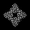
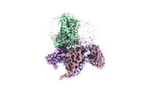
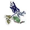
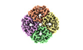
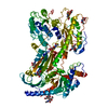
EMDB-36635: 
Structure of arginine oxidase from Pseudomonas sp. TRU 7192
PDB-8jt7:
Structure of arginine oxidase from Pseudomonas sp. TRU 7192
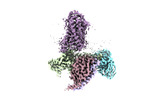
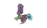
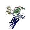
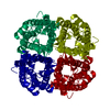
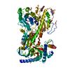
EMDB-35442:
Cryo-EM structure of HCA2-Gi complex with GSK256073
PDB-8ihb:
Cryo-EM structure of HCA2-Gi complex with GSK256073

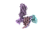
EMDB-35443:
Cryo-EM structure of HCA2-Gi complex with MK6892
EMDB-35444:
Cryo-EM structure of HCA2-Gi complex with LUF6283
EMDB-35445:
Cryo-EM structure of HCA2-Gi complex with acifran
EMDB-35446:
Cryo-EM structure of HCA3-Gi complex with acifran
EMDB-35447:
Cryo-EM structure of HCA3-Gi complex with acifran (local)
PDB-8ihf:
Cryo-EM structure of HCA2-Gi complex with MK6892
PDB-8ihh:
Cryo-EM structure of HCA2-Gi complex with LUF6283

PDB-8ihi:
Cryo-EM structure of HCA2-Gi complex with acifran
PDB-8ihj:
Cryo-EM structure of HCA3-Gi complex with acifran
PDB-8ihk:
Cryo-EM structure of HCA3-Gi complex with acifran (local)
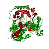
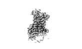
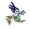
EMDB-29934:
Cryo-EM structure of hAQP2 in DDM
PDB-8gcl:
Cryo-EM structure of hAQP2 in DDM
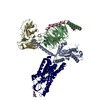
EMDB-35399:
Structure of human alpha-2/delta-1 with mirogabalin
EMDB-35400:
Structure of human alpha-2/delta-1 without mirogabalin
PDB-8if3:
Structure of human alpha-2/delta-1 with mirogabalin
PDB-8if4:
Structure of human alpha-2/delta-1 without mirogabalin

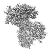
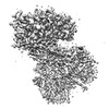
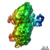
EMDB-0893:
Detailed structures of the gliding machinery observed by ECT
EMDB-5931:
Dictyostelium dynein microtubule binding domain bound to a 15-protofilament microtubule

PDB-3j6p:
Pseudo-atomic model of dynein microtubule binding domain-tubulin complex based on a cryoEM map
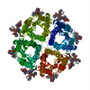
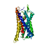
PDB-2zz9:
Structure of aquaporin-4 S180D mutant at 2.8 A resolution by electron crystallography

PDB-2d57:
Double layered 2D crystal structure of AQUAPORIN-4 (AQP4M23) at 3.2 a resolution by electron crystallography
 wwPDBはEMDBデータモデルのバージョン3へ移行します
