 Movie
Movie Controller


+ Open data

- Basic information
| Entry | Database: PDB / ID: 8jt7 | ||||||
|---|---|---|---|---|---|---|---|
| Title | Structure of arginine oxidase from Pseudomonas sp. TRU 7192 | ||||||
 Components Components | Amine oxidoreductase | ||||||
 Keywords Keywords | OXIDOREDUCTASE | ||||||
| Function / homology | FLAVIN-ADENINE DINUCLEOTIDE Function and homology information Function and homology information | ||||||
| Biological species |  Pseudomonas sp. (bacteria) Pseudomonas sp. (bacteria) | ||||||
| Method | ELECTRON MICROSCOPY / single particle reconstruction / cryo EM / Resolution: 2.34 Å | ||||||
 Authors Authors | Yamaguchi, H. / Numoto, N. / Suzuki, H. / Nishikawa, K. / Kamegawa, A. / Takahashi, K. / Sugiki, M. / Fujiyoshi, Y. | ||||||
| Funding support | 1items
| ||||||
 Citation Citation | Journal: J Biochem / Year: 2025 Title: Open and closed structures of L-arginine oxidase by cryo-electron microscopy and X-ray crystallography. Authors: Hiroki Yamaguchi / Kazutoshi Takahashi / Nobutaka Numoto / Hiroshi Suzuki / Moemi Tatsumi / Akiko Kamegawa / Kouki Nishikawa / Yasuhisa Asano / Toshimi Mizukoshi / Hiroshi Miyano / Yoshinori ...Authors: Hiroki Yamaguchi / Kazutoshi Takahashi / Nobutaka Numoto / Hiroshi Suzuki / Moemi Tatsumi / Akiko Kamegawa / Kouki Nishikawa / Yasuhisa Asano / Toshimi Mizukoshi / Hiroshi Miyano / Yoshinori Fujiyoshi / Masayuki Sugiki /  Abstract: L-arginine oxidase (AROD, EC 1.4.3.25) is an oxidoreductase that catalyses the deamination of L-arginine, with flavin adenine dinucleotide (FAD) as a cofactor. Recently identified AROD from ...L-arginine oxidase (AROD, EC 1.4.3.25) is an oxidoreductase that catalyses the deamination of L-arginine, with flavin adenine dinucleotide (FAD) as a cofactor. Recently identified AROD from Pseudomonas sp. TPU 7192 (PT-AROD) demonstrates high selectivity for L-arginine. This enzyme is useful for accurate assays of L-arginine in biological samples. The structural characteristics of the FAD-dependent AROD, however, remain unknown. Here, we report the structure of PT-AROD at a resolution of 2.3 Å by cryo-electron microscopy. PT-AROD adopts an octameric structure with D4 symmetry, which is consistent with its molecular weight in solution, estimated by mass photometry. Comparative analysis of this structure with that determined using X-ray crystallography reveals open and closed forms of the lid-like loop at the entrance to the substrate pocket. Furthermore, mutation of Glu493, located at the substrate binding site, diminishes substrate selectivity, suggesting that this residue contributes significantly to the high selectivity of PT-AROD. | ||||||
| History |
|
- Structure visualization
| Structure viewer | Molecule: MolmilJmol/JSmol |
|---|
- Downloads & links
-Download
| PDBx/mmCIF format | 8jt7.cif.gz | 129.4 KB | Display | PDBx/mmCIF format |
|---|---|---|---|---|
| PDB format | pdb8jt7.ent.gz | 98.6 KB | Display | PDB format |
| PDBx/mmJSON format | 8jt7.json.gz | Tree view | PDBx/mmJSON format | |
| Others |  Other downloads Other downloads |
-Validation report
| Arichive directory | https://data.pdbj.org/pub/pdb/validation_reports/jt/8jt7ftp://data.pdbj.org/pub/pdb/validation_reports/jt/8jt7 | HTTPS FTP |
|---|
-Related structure data
| Related structure data |  36635MC  8t8aC M: map data used to model this data C: citing same article ( |
|---|---|
| Similar structure data |
-Links
 PDBj
PDBj- Assembly
| Deposited unit | 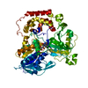
| ||||||||||||||||||||||||||||||||
|---|---|---|---|---|---|---|---|---|---|---|---|---|---|---|---|---|---|---|---|---|---|---|---|---|---|---|---|---|---|---|---|---|---|
| 1 | x 8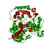
| ||||||||||||||||||||||||||||||||
| Noncrystallographic symmetry (NCS) | NCS oper:
|
-Components
| #1: Protein | Mass: 67393.164 Da / Num. of mol.: 1 / Fragment: initiation methionine (1A), his-tags (2-7A) Source method: isolated from a genetically manipulated source Source: (gene. exp.) Pseudomonas sp. (bacteria) / Strain: TPU 7192 / Production host: |
|---|---|
| #2: Chemical | ChemComp-FAD /   Mass: 785.550 Da / Num. of mol.: 1 / Source method: isolated from a natural source / Formula: C27H33N9O15P2 / Feature type: SUBJECT OF INVESTIGATION / Comment: FAD*YM Mass: 785.550 Da / Num. of mol.: 1 / Source method: isolated from a natural source / Formula: C27H33N9O15P2 / Feature type: SUBJECT OF INVESTIGATION / Comment: FAD*YM |
| #3: Water | ChemComp-HOH /  Mass: 18.015 Da / Num. of mol.: 29 / Source method: isolated from a natural source / Formula: H2O Mass: 18.015 Da / Num. of mol.: 29 / Source method: isolated from a natural source / Formula: H2O |
| Has ligand of interest | Y |
| Has protein modification | N |
-Experimental details
-Experiment
| Experiment | Method: ELECTRON MICROSCOPY |
|---|---|
| EM experiment | Aggregation state: PARTICLE / 3D reconstruction method: single particle reconstruction |
- Sample preparation
| Component | Name: homooctamer of arginine oxidase / Type: COMPLEX / Entity ID: #1 / Source: RECOMBINANT | |||||||||||||||
|---|---|---|---|---|---|---|---|---|---|---|---|---|---|---|---|---|
| Molecular weight | Value: 0.54 MDa / Experimental value: NO | |||||||||||||||
| Source (natural) | Organism: Pseudomonas sp. (bacteria) / Strain: TPU 7192 | |||||||||||||||
| Source (recombinant) | Organism: | |||||||||||||||
| Buffer solution | pH: 8 | |||||||||||||||
| Buffer component |
| |||||||||||||||
| Specimen | Conc.: 2.5 mg/ml / Embedding applied: NO / Shadowing applied: NO / Staining applied: NO / Vitrification applied: YES | |||||||||||||||
| Specimen support | Grid material: MOLYBDENUM / Grid mesh size: 200 divisions/in. / Grid type: Quantifoil R2/2 | |||||||||||||||
| Vitrification | Instrument: LEICA KF80 / Cryogen name: ETHANE |
- Electron microscopy imaging
| Microscopy | Model: JEOL CRYO ARM 300 |
|---|---|
| Electron gun | Electron source:  FIELD EMISSION GUN / Accelerating voltage: 300 kV / Illumination mode: FLOOD BEAM FIELD EMISSION GUN / Accelerating voltage: 300 kV / Illumination mode: FLOOD BEAM |
| Electron lens | Mode: BRIGHT FIELD / Nominal defocus max: 2000 nm / Nominal defocus min: 1000 nm / Cs: 3.4 mm / C2 aperture diameter: 50 µm / Alignment procedure: COMA FREE |
| Specimen holder | Cryogen: NITROGEN |
| Image recording | Average exposure time: 8 sec. / Electron dose: 69.6 e/Å2 / Detector mode: COUNTING / Film or detector model: GATAN K2 SUMMIT (4k x 4k) / Num. of grids imaged: 1 / Num. of real images: 5035 |
| EM imaging optics | Energyfilter name: In-column Omega Filter / Energyfilter slit width: 20 eV |
| Image scans | Movie frames/image: 40 |
- Processing
| EM software |
| ||||||||||||||||||||||||||||||||||||||||
|---|---|---|---|---|---|---|---|---|---|---|---|---|---|---|---|---|---|---|---|---|---|---|---|---|---|---|---|---|---|---|---|---|---|---|---|---|---|---|---|---|---|
| CTF correction | Type: PHASE FLIPPING AND AMPLITUDE CORRECTION | ||||||||||||||||||||||||||||||||||||||||
| Symmetry | Point symmetry: D4 (2x4 fold dihedral) | ||||||||||||||||||||||||||||||||||||||||
| 3D reconstruction | Resolution: 2.34 Å / Resolution method: FSC 0.143 CUT-OFF / Num. of particles: 254111 / Symmetry type: POINT | ||||||||||||||||||||||||||||||||||||||||
| Atomic model building | Source name: AlphaFold / Type: in silico model |
