 Movie
Movie Controller


+ Open data

- Basic information
| Entry | Database: PDB / ID: 8ihj | |||||||||||||||||||||||||||||||||||||||||||||||||||||||||||||||
|---|---|---|---|---|---|---|---|---|---|---|---|---|---|---|---|---|---|---|---|---|---|---|---|---|---|---|---|---|---|---|---|---|---|---|---|---|---|---|---|---|---|---|---|---|---|---|---|---|---|---|---|---|---|---|---|---|---|---|---|---|---|---|---|---|
| Title | Cryo-EM structure of HCA3-Gi complex with acifran | |||||||||||||||||||||||||||||||||||||||||||||||||||||||||||||||
 Components Components |
| |||||||||||||||||||||||||||||||||||||||||||||||||||||||||||||||
 Keywords Keywords | SIGNALING PROTEIN / GPCR | |||||||||||||||||||||||||||||||||||||||||||||||||||||||||||||||
| Function / homology |  Function and homology information Function and homology informationActivation of the phototransduction cascade / Olfactory Signaling Pathway / Synthesis, secretion, and inactivation of Glucagon-like Peptide-1 (GLP-1) / Sensory perception of sweet, bitter, and umami (glutamate) taste / G beta:gamma signalling through PLC beta / Presynaptic function of Kainate receptors / Prostacyclin signalling through prostacyclin receptor / G alpha (z) signalling events / Glucagon-type ligand receptors / G beta:gamma signalling through PI3Kgamma ...Activation of the phototransduction cascade / Olfactory Signaling Pathway / Synthesis, secretion, and inactivation of Glucagon-like Peptide-1 (GLP-1) / Sensory perception of sweet, bitter, and umami (glutamate) taste / G beta:gamma signalling through PLC beta / Presynaptic function of Kainate receptors / Prostacyclin signalling through prostacyclin receptor / G alpha (z) signalling events / Glucagon-type ligand receptors / G beta:gamma signalling through PI3Kgamma / G beta:gamma signalling through CDC42 / Adrenaline,noradrenaline inhibits insulin secretion / ADP signalling through P2Y purinoceptor 12 / Cooperation of PDCL (PhLP1) and TRiC/CCT in G-protein beta folding / Thromboxane signalling through TP receptor / G beta:gamma signalling through BTK / Thrombin signalling through proteinase activated receptors (PARs) / Activation of G protein gated Potassium channels / Inhibition of voltage gated Ca2+ channels via Gbeta/gamma subunits / G alpha (s) signalling events / Ca2+ pathway / G-protein activation / nicotinic acid receptor activity / Extra-nuclear estrogen signaling / G alpha (12/13) signalling events / Hydroxycarboxylic acid-binding receptors / G alpha (q) signalling events / Vasopressin regulates renal water homeostasis via Aquaporins / GPER1 signaling / Glucagon-like Peptide-1 (GLP1) regulates insulin secretion / ADP signalling through P2Y purinoceptor 1 / High laminar flow shear stress activates signaling by PIEZO1 and PECAM1:CDH5:KDR in endothelial cells / G alpha (i) signalling events / photoreceptor outer segment membrane / spectrin binding / alkylglycerophosphoethanolamine phosphodiesterase activity / phototransduction, visible light / photoreceptor outer segment / negative regulation of lipid catabolic process / regulation of eating behavior / cardiac muscle cell apoptotic process / adenylate cyclase inhibitor activity / photoreceptor inner segment / positive regulation of protein localization to cell cortex / T cell migration / positive regulation of relaxation of smooth muscle / Adenylate cyclase inhibitory pathway / D2 dopamine receptor binding / adenylate cyclase-inhibiting serotonin receptor signaling pathway / G protein-coupled serotonin receptor binding / cellular response to forskolin / mast cell degranulation / regulation of mitotic spindle organization / chemokine-mediated signaling pathway / Regulation of insulin secretion / neuropeptide signaling pathway / response to prostaglandin E / electron transport chain / positive regulation of cholesterol biosynthetic process / G protein-coupled receptor binding / response to peptide hormone / G protein-coupled receptor activity / G-protein beta/gamma-subunit complex binding / adenylate cyclase-modulating G protein-coupled receptor signaling pathway / cell junction / adenylate cyclase-inhibiting G protein-coupled receptor signaling pathway / GDP binding / ADP signalling through P2Y purinoceptor 12 / Adrenaline,noradrenaline inhibits insulin secretion / myelin sheath / G alpha (z) signalling events / ADORA2B mediated anti-inflammatory cytokines production / GPER1 signaling / heterotrimeric G-protein complex / G-protein beta-subunit binding / positive regulation of cytosolic calcium ion concentration / sensory perception of taste / signaling receptor complex adaptor activity / adenylate cyclase-activating G protein-coupled receptor signaling pathway / retina development in camera-type eye / cell body / GTPase binding / G protein activity / midbody / cell cortex / cellular response to hypoxia / G alpha (i) signalling events / G alpha (s) signalling events / phospholipase C-activating G protein-coupled receptor signaling pathway / Hydrolases; Acting on acid anhydrides; Acting on GTP to facilitate cellular and subcellular movement / periplasmic space / electron transfer activity / cell population proliferation / Extra-nuclear estrogen signaling / ciliary basal body / iron ion binding / G protein-coupled receptor signaling pathway / cell division / lysosomal membrane / GTPase activity Similarity search - Function | |||||||||||||||||||||||||||||||||||||||||||||||||||||||||||||||
| Biological species |   Homo sapiens (human) Homo sapiens (human)synthetic construct (others)  | |||||||||||||||||||||||||||||||||||||||||||||||||||||||||||||||
| Method | ELECTRON MICROSCOPY / single particle reconstruction / cryo EM / Resolution: 3.07 Å | |||||||||||||||||||||||||||||||||||||||||||||||||||||||||||||||
 Authors Authors | Suzuki, S. / Nishikawa, K. / Suzuki, H. / Fujiyoshi, Y. | |||||||||||||||||||||||||||||||||||||||||||||||||||||||||||||||
| Funding support |  Japan, 1items Japan, 1items
| |||||||||||||||||||||||||||||||||||||||||||||||||||||||||||||||
 Citation Citation | Journal: Nat Commun / Year: 2023 Title: Structural basis of hydroxycarboxylic acid receptor signaling mechanisms through ligand binding. Authors: Shota Suzuki / Kotaro Tanaka / Kouki Nishikawa / Hiroshi Suzuki / Atsunori Oshima / Yoshinori Fujiyoshi / Abstract: Hydroxycarboxylic acid receptors (HCA) are expressed in various tissues and immune cells. HCA2 and its agonist are thus important targets for treating inflammatory and metabolic disorders. Only ...Hydroxycarboxylic acid receptors (HCA) are expressed in various tissues and immune cells. HCA2 and its agonist are thus important targets for treating inflammatory and metabolic disorders. Only limited information is available, however, on the active-state binding of HCAs with agonists. Here, we present cryo-EM structures of human HCA2-Gi and HCA3-Gi signaling complexes binding with multiple compounds bound. Agonists were revealed to form a salt bridge with arginine, which is conserved in the HCA family, to activate these receptors. Extracellular regions of the receptors form a lid-like structure that covers the ligand-binding pocket. Although transmembrane (TM) 6 in HCAs undergoes dynamic conformational changes, ligands do not directly interact with amino acids in TM6, suggesting that indirect signaling induces a slight shift in TM6 to activate Gi proteins. Structural analyses of agonist-bound HCA2 and HCA3 together with mutagenesis and molecular dynamics simulation provide molecular insights into HCA ligand recognition and activation mechanisms. | |||||||||||||||||||||||||||||||||||||||||||||||||||||||||||||||
| History |
|
- Structure visualization
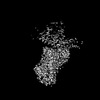
| Structure viewer | Molecule: MolmilJmol/JSmol |
|---|
- Downloads & links
-Download
| PDBx/mmCIF format | 8ihj.cif.gz | 240.7 KB | Display | PDBx/mmCIF format |
|---|---|---|---|---|
| PDB format | pdb8ihj.ent.gz | 179.3 KB | Display | PDB format |
| PDBx/mmJSON format | 8ihj.json.gz | Tree view | PDBx/mmJSON format | |
| Others |  Other downloads Other downloads |
-Validation report
| Arichive directory | https://data.pdbj.org/pub/pdb/validation_reports/ih/8ihjftp://data.pdbj.org/pub/pdb/validation_reports/ih/8ihj | HTTPS FTP |
|---|
-Related structure data
| Related structure data | 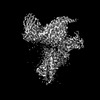 35446MC 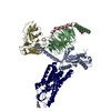 8ihbC 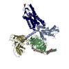 8ihfC 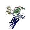 8ihhC  8ihiC 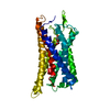 8ihkC M: map data used to model this data C: citing same article ( |
|---|---|
| Similar structure data |

-Links
 PDBj
PDBj


- Assembly
| Deposited unit | 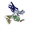
|
|---|---|
| 1 |
|
-Components
-Guanine nucleotide-binding protein ... , 3 types, 3 molecules ABC
| #2: Protein | Mass: 40446.047 Da / Num. of mol.: 1 / Mutation: G203A, A326S Source method: isolated from a genetically manipulated source Source: (gene. exp.) Homo sapiens (human) / Gene: GNAI1 / Production host:   Spodoptera frugiperda (fall armyworm) / References: UniProt: P63096 Spodoptera frugiperda (fall armyworm) / References: UniProt: P63096 |
|---|---|
| #4: Protein | Mass: 41772.562 Da / Num. of mol.: 1 Source method: isolated from a genetically manipulated source Source: (gene. exp.) Spodoptera frugiperda (fall armyworm) / References: UniProt: P62874 |
| #5: Protein | Mass: 7729.947 Da / Num. of mol.: 1 Source method: isolated from a genetically manipulated source Source: (gene. exp.) Spodoptera frugiperda (fall armyworm) / References: UniProt: P63213 |
-Protein / Antibody / Non-polymers , 3 types, 3 molecules RS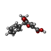
| #1: Protein | Mass: 60749.406 Da / Num. of mol.: 1 / Mutation: M29W,H124I Source method: isolated from a genetically manipulated source Source: (gene. exp.) Homo sapiens (human)Gene: cybC, HCAR3, GPR109B, HCA3, HM74B, NIACR2 / Production host: Spodoptera frugiperda (fall armyworm) / References: UniProt: P0ABE7, UniProt: P49019 |
|---|---|
| #3: Antibody | Mass: 26496.514 Da / Num. of mol.: 1 Source method: isolated from a genetically manipulated source Source: (gene. exp.) synthetic construct (others) / Production host: Spodoptera frugiperda (fall armyworm) |
| #6: Chemical | ChemComp-P9X / ( Mass: 218.205 Da / Num. of mol.: 1 / Source method: obtained synthetically / Formula: C12H10O4 / Feature type: SUBJECT OF INVESTIGATION Mass: 218.205 Da / Num. of mol.: 1 / Source method: obtained synthetically / Formula: C12H10O4 / Feature type: SUBJECT OF INVESTIGATION |
-Details
| Has ligand of interest | Y |
|---|---|
| Has protein modification | Y |
-Experimental details
-Experiment
| Experiment | Method: ELECTRON MICROSCOPY |
|---|---|
| EM experiment | Aggregation state: PARTICLE / 3D reconstruction method: single particle reconstruction |
- Sample preparation
| Component | Name: Multiprotein complex / Type: COMPLEX / Entity ID: #1-#2, #4-#5, #3 / Source: MULTIPLE SOURCES |
|---|---|
| Molecular weight | Experimental value: NO |
| Source (natural) | Organism: Homo sapiens (human) |
| Source (recombinant) | Organism: Spodoptera frugiperda (fall armyworm) |
| Buffer solution | pH: 7.4 |
| Specimen | Conc.: 15 mg/ml / Embedding applied: NO / Shadowing applied: NO / Staining applied: NO / Vitrification applied: YES / Details: This sample was monodisperse |
| Vitrification | Instrument: FEI VITROBOT MARK IV / Cryogen name: ETHANE / Humidity: 100 % / Chamber temperature: 298 K |
- Electron microscopy imaging
| Microscopy | Model: JEOL CRYO ARM 300 |
|---|---|
| Electron gun | Electron source:  FIELD EMISSION GUN / Accelerating voltage: 300 kV / Illumination mode: FLOOD BEAM FIELD EMISSION GUN / Accelerating voltage: 300 kV / Illumination mode: FLOOD BEAM |
| Electron lens | Mode: BRIGHT FIELD / Nominal defocus max: 2000 nm / Nominal defocus min: 1000 nm |
| Image recording | Electron dose: 49 e/Å2 / Film or detector model: GATAN K3 (6k x 4k) |
- Processing
| EM software | Name: PHENIX / Category: model refinement | ||||||||||||||||||||||||
|---|---|---|---|---|---|---|---|---|---|---|---|---|---|---|---|---|---|---|---|---|---|---|---|---|---|
| CTF correction | Type: PHASE FLIPPING AND AMPLITUDE CORRECTION | ||||||||||||||||||||||||
| 3D reconstruction | Resolution: 3.07 Å / Resolution method: FSC 0.143 CUT-OFF / Num. of particles: 95567 / Symmetry type: POINT | ||||||||||||||||||||||||
| Refine LS restraints |
|
