 Movie
Movie Controller
 Structure viewers
 About EMN search


-Search query
-Search result
Showing 1 - 50 of 85 items for (author: maud & d)

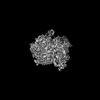
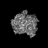
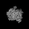
EMDB-53306: 
GH161 phosphorylase in complex with beta-1,3 glucooligosaccharide
Method: single particle / : Cooper N, Cioci G, Ladeveze S, Potocki-Veronese G, Moulis C, Remaud-Simeon M

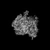
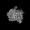
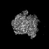
EMDB-55127:
Catalytically active GH161A phosphorylase refined in C1
Method: single particle / : Cooper N, Cioci G, Ladeveze S, Potocki-Veronese G, Moulis C

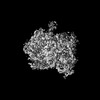
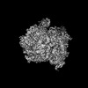
PDB-9qqk:
GH161 phosphorylase in complex with beta-1,3 glucooligosaccharide
Method: single particle / : Cooper N, Cioci G, Ladeveze S, Potocki-Veronese G, Moulis C, Remaud-Simeon M

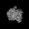
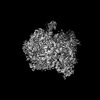
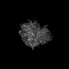
EMDB-51944:
The Cryo-EM structure of bacterial beta-1,3-glucan phosphorylase from family GH161
Method: single particle / : Cioci G, Cooper N, Ladeveze S, Shayan R

EMDB-50892:
Influenza A/H7N9 polymerase pre-cleavage cap-snatching complex
Method: single particle / : Rotsch AH, Li D, Dienemann C, Cusack S, Cramer P

EMDB-50927:
Influenza A/H7N9 polymerase post-cleavage cap-snatching complex
Method: single particle / : Rotsch AH, Li D, Dienemann C, Cusack S, Cramer P

PDB-9fyx:
Influenza A/H7N9 polymerase pre-cleavage cap-snatching complex
Method: single particle / : Rotsch AH, Li D, Dienemann C, Cusack S, Cramer P

PDB-9g0a:
Influenza A/H7N9 polymerase post-cleavage cap-snatching complex
Method: single particle / : Rotsch AH, Li D, Dienemann C, Cusack S, Cramer P
EMDB-50675:
Escherichia coli 70S ribosome in situ structure
Method: subtomogram averaging / : Watson H, Berger C, Grange M
EMDB-50676:
E. coli 70S ribosome in situ structure for xenon damage layer determination: 5 - 10 nm
Method: subtomogram averaging / : Watson H, Berger C, Grange M
EMDB-50678:
E. coli 70S ribosome in situ structure for xenon damage layer determination: 10 - 15 nm
Method: subtomogram averaging / : Watson H, Berger C, Grange M
EMDB-50679:
E. coli 70S ribosome in situ structure for xenon damage layer determination: 15 - 20 nm
Method: subtomogram averaging / : Watson H, Berger C, Grange M

EMDB-50680:
E. coli 70S ribosome in situ structure for xenon damage layer determination: 20 - 25 nm
Method: subtomogram averaging / : Watson H, Berger C, Grange M

EMDB-50681:
E. coli 70S ribosome in situ structure for xenon damage layer determination: 25 - 30 nm
Method: subtomogram averaging / : Watson H, Berger C, Grange M
EMDB-50682:
E. coli 70S ribosome in situ structure for xenon damage layer determination: 30 - 35 nm
Method: subtomogram averaging / : Watson H, Berger C, Grange M
EMDB-50683:
E. coli 70S ribosome in situ structure for xenon damage layer determination: 35 - 40 nm
Method: subtomogram averaging / : Watson H, Berger C, Grange M
EMDB-50684:
E. coli 70S ribosome in situ structure for xenon damage layer determination: 40 - 45 nm
Method: subtomogram averaging / : Watson H, Berger C, Grange M
EMDB-50685:
E. coli 70S ribosome in situ structure for xenon damage layer determination: 45 - 50 nm
Method: subtomogram averaging / : Watson H, Berger C, Grange M
EMDB-50686:
E. coli 70S ribosome in situ structure for xenon damage layer determination: 50 - 55 nm
Method: subtomogram averaging / : Watson H, Berger C, Grange M

EMDB-50687:
E. coli 70S ribosome in situ structure for xenon damage layer determination: 55 - 60 nm
Method: subtomogram averaging / : Watson H, Berger C, Grange M
EMDB-50688:
E. coli 70S ribosome in situ structure for xenon damage layer determination: >10nm matched control for 5 - 10 nm
Method: subtomogram averaging / : Watson H, Berger C, Grange M

EMDB-50689:
E. coli 70S ribosome in situ structure for xenon damage layer determination: >15 nm matched control for 10 - 15 nm
Method: subtomogram averaging / : Watson H, Berger C, Grange M

EMDB-50690:
E. coli 70S ribosome in situ structure for xenon damage layer determination: >20 nm matched control for 15 - 20 nm
Method: subtomogram averaging / : Watson H, Berger C, Grange M

EMDB-50691:
E. coli 70S ribosome in situ structure for xenon damage layer determination: >25 nm matched control for 20 - 25 nm
Method: subtomogram averaging / : Watson H, Berger C, Grange M

EMDB-50692:
E. coli 70S ribosome in situ structure for xenon damage layer determination: >30 nm matched control for 25 - 30 nm
Method: subtomogram averaging / : Watson H, Berger C, Grange M

EMDB-50693:
E. coli 70S ribosome in situ structure for xenon damage layer determination: >35 nm matched control for 30 - 35 nm
Method: subtomogram averaging / : Watson H, Berger C, Grange M

EMDB-50694:
E. coli 70S ribosome in situ structure for xenon damage layer determination: >40 nm matched control for 35 - 40 nm
Method: subtomogram averaging / : Watson H, Berger C, Grange M

EMDB-50695:
E. coli 70S ribosome in situ structure for xenon damage layer determination: >45 nm matched control for 40 - 45 nm
Method: subtomogram averaging / : Watson H, Berger C, Grange M

EMDB-50696:
E. coli 70S ribosome in situ structure for xenon damage layer determination: >50 nm matched control for 45 - 50 nm
Method: subtomogram averaging / : Watson H, Berger C, Grange M

EMDB-50697:
E. coli 70S ribosome in situ structure for xenon damage layer determination: >55 nm matched control for 50 - 55 nm
Method: subtomogram averaging / : Watson H, Berger C, Grange M

EMDB-50698:
E. coli 70S ribosome in situ structure for xenon damage layer determination: >60 nm matched control for 55 - 60 nm
Method: subtomogram averaging / : Watson H, Berger C, Grange M

EMDB-50699:
E. coli 70S ribosome in situ structure for lamellae backside damage analysis: 0 - 1 micron from lamellae backside
Method: subtomogram averaging / : Watson H, Berger C, Grange M

EMDB-50700:
E. coli 70S ribosome in situ structure for lamellae backside damage analysis: 1 - 2 microns from lamellae backside
Method: subtomogram averaging / : Watson H, Berger C, Grange M

EMDB-50701:
E. coli 70S ribosome in situ structure for lamellae backside damage analysis: 2 - 3 microns from lamellae backside
Method: subtomogram averaging / : Watson H, Berger C, Grange M

EMDB-50702:
E. coli 70S ribosome in situ structure for lamellae backside damage analysis: 3 - 4 microns from lamellae backside
Method: subtomogram averaging / : Watson H, Berger C, Grange M

EMDB-50703:
E. coli 70S ribosome in situ structure for lamellae backside damage analysis: 4 - 5 microns from lamellae backside
Method: subtomogram averaging / : Watson H, Berger C, Grange M

EMDB-50704:
E. coli 70S ribosome in situ structure for xenon damage layer determination: 10,000 particles (no depth constraint)
Method: subtomogram averaging / : Watson H, Berger C, Grange M

EMDB-50705:
E. coli 70S ribosome in situ structure for xenon damage layer determination: 10,000 particles (>45 nm)
Method: subtomogram averaging / : Watson H, Berger C, Grange M

EMDB-50706:
E. coli 70S ribosome in situ structure for xenon damage layer determination: 10,000 particles (<=45 nm)
Method: subtomogram averaging / : Watson H, Berger C, Grange M

EMDB-52177:
E. coli 70S ribosome in situ structure for xenon damage layer determination: 10,000 particles (no depth constraint)
Method: subtomogram averaging / : Watson H, Berger C, Grange M

EMDB-52178:
E. coli 70S ribosome in situ structure for xenon damage layer determination: 10,000 particles (<30 nm)
Method: subtomogram averaging / : Watson H, Berger C, Grange M

EMDB-52179:
E. coli 70S ribosome in situ structure for xenon damage layer determination: 10,000 particles (>30 nm)
Method: subtomogram averaging / : Watson H, Berger C, Grange M

EMDB-52438:
In-cell Structure of Pyrenoid Rubisco
Method: subtomogram averaging / : Nadav E, Zhen H, Maud D, Alireza R, Juan R P, Peijun P

PDB-9hvm:
In-cell Structure of Pyrenoid Rubisco
Method: subtomogram averaging / : Nadav E, Zhen H, Maud D, Alireza R, Juan R P, Peijun P

EMDB-15525:
Cryo-EM structure of the RecA postsynaptic filament from S. pneumoniae
Method: helical / : Perry TN, Fronzes R, Polard P, Hertzog M

PDB-8amf:
Cryo-EM structure of the RecA postsynaptic filament from S. pneumoniae
Method: helical / : Perry TN, Fronzes R, Polard P, Hertzog M

EMDB-17767:
Cryo electron tomography of human choriocarcinoma cells
Method: electron tomography / : Tun WM, Yee NB-Y, Ho EML, Darrow MC, Basham M

EMDB-15524:
Cryo-EM structure of the RecA presynaptic filament from S.pneumoniae
Method: helical / : Perry TN, Fronzes R, Polard P, Hertzog M

PDB-8amd:
Cryo-EM structure of the RecA presynaptic filament from S.pneumoniae
Method: helical / : Perry TN, Fronzes R, Polard P, Hertzog M
EMDB-15636:
Human 80S ribosome structure from pFIB-lamellae
Method: subtomogram averaging / : Berger C, Grange M
Pages:
 wwPDB to switch to version 3 of the EMDB data model
