 Movie
Movie Controller
 Structure viewers
 About Yorodumi Papers


+Search query
-Structure paper
| Title | Xenon plasma focused ion beam lamella fabrication on high-pressure frozen specimens for structural cell biology. |
|---|---|
| Journal, issue, pages | Nat Commun, Vol. 16, Issue 1, Page 2286, Year 2025 |
| Publish date | Mar 7, 2025 |
 Authors Authors | Casper Berger / Helena Watson / James H Naismith / Maud Dumoux / Michael Grange /  |
| PubMed Abstract | Cryo focused ion beam lamella preparation is a potent tool for in situ structural biology, enabling the study of macromolecules in their native cellular environments. However, throughput is currently ...Cryo focused ion beam lamella preparation is a potent tool for in situ structural biology, enabling the study of macromolecules in their native cellular environments. However, throughput is currently limited, especially for thicker, more biologically complex samples. We describe how xenon plasma focused ion beam milling can be used for routine bulk milling of thicker, high-pressure frozen samples. We demonstrate lamellae preparation with a high success rate on these samples and determine a 4.0 Å structure of the Escherichia coli ribosome on these lamellae using sub volume averaging. We determine the effects on sample integrity of increased ion currents up to 60 nA during bulk milling of thicker planar samples, showing no measurable damage to macromolecules beyond an amorphous layer on the backside of the lamellae. The use of xenon results in substantial structural damage to particles up to approximately 30 nm in depth from the milled surfaces, and the effects of damage become negligibly small by 45 nm. Our results outline how the use of high currents using xenon plasma focused ion beam milling may be integrated into FIB milling regimes for preparing thin lamellae for high-resolution in situ structural biology. |
 External links External links | Nat Commun / PubMed:40055361 / PubMed Central |
| Methods | EM (subtomogram averaging) |
| Resolution | 4.0 - 15.8 Å |
| Structure data |  EMDB-50675: Escherichia coli 70S ribosome in situ structure  EMDB-50676: E. coli 70S ribosome in situ structure for xenon damage layer determination: 5 - 10 nm  EMDB-50678: E. coli 70S ribosome in situ structure for xenon damage layer determination: 10 - 15 nm  EMDB-50679: E. coli 70S ribosome in situ structure for xenon damage layer determination: 15 - 20 nm  EMDB-50680: E. coli 70S ribosome in situ structure for xenon damage layer determination: 20 - 25 nm  EMDB-50681: E. coli 70S ribosome in situ structure for xenon damage layer determination: 25 - 30 nm  EMDB-50682: E. coli 70S ribosome in situ structure for xenon damage layer determination: 30 - 35 nm  EMDB-50683: E. coli 70S ribosome in situ structure for xenon damage layer determination: 35 - 40 nm  EMDB-50684: E. coli 70S ribosome in situ structure for xenon damage layer determination: 40 - 45 nm  EMDB-50685: E. coli 70S ribosome in situ structure for xenon damage layer determination: 45 - 50 nm 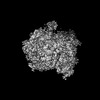 EMDB-50686: E. coli 70S ribosome in situ structure for xenon damage layer determination: 50 - 55 nm  EMDB-50687: E. coli 70S ribosome in situ structure for xenon damage layer determination: 55 - 60 nm 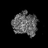 EMDB-50688: E. coli 70S ribosome in situ structure for xenon damage layer determination: >10nm matched control for 5 - 10 nm  EMDB-50689: E. coli 70S ribosome in situ structure for xenon damage layer determination: >15 nm matched control for 10 - 15 nm  EMDB-50690: E. coli 70S ribosome in situ structure for xenon damage layer determination: >20 nm matched control for 15 - 20 nm  EMDB-50691: E. coli 70S ribosome in situ structure for xenon damage layer determination: >25 nm matched control for 20 - 25 nm  EMDB-50692: E. coli 70S ribosome in situ structure for xenon damage layer determination: >30 nm matched control for 25 - 30 nm  EMDB-50693: E. coli 70S ribosome in situ structure for xenon damage layer determination: >35 nm matched control for 30 - 35 nm  EMDB-50694: E. coli 70S ribosome in situ structure for xenon damage layer determination: >40 nm matched control for 35 - 40 nm  EMDB-50695: E. coli 70S ribosome in situ structure for xenon damage layer determination: >45 nm matched control for 40 - 45 nm  EMDB-50696: E. coli 70S ribosome in situ structure for xenon damage layer determination: >50 nm matched control for 45 - 50 nm  EMDB-50697: E. coli 70S ribosome in situ structure for xenon damage layer determination: >55 nm matched control for 50 - 55 nm  EMDB-50698: E. coli 70S ribosome in situ structure for xenon damage layer determination: >60 nm matched control for 55 - 60 nm  EMDB-50699: E. coli 70S ribosome in situ structure for lamellae backside damage analysis: 0 - 1 micron from lamellae backside  EMDB-50700: E. coli 70S ribosome in situ structure for lamellae backside damage analysis: 1 - 2 microns from lamellae backside  EMDB-50701: E. coli 70S ribosome in situ structure for lamellae backside damage analysis: 2 - 3 microns from lamellae backside  EMDB-50702: E. coli 70S ribosome in situ structure for lamellae backside damage analysis: 3 - 4 microns from lamellae backside  EMDB-50703: E. coli 70S ribosome in situ structure for lamellae backside damage analysis: 4 - 5 microns from lamellae backside  EMDB-50704: E. coli 70S ribosome in situ structure for xenon damage layer determination: 10,000 particles (no depth constraint)  EMDB-50705: E. coli 70S ribosome in situ structure for xenon damage layer determination: 10,000 particles (>45 nm)  EMDB-50706: E. coli 70S ribosome in situ structure for xenon damage layer determination: 10,000 particles (<=45 nm)  EMDB-52177: E. coli 70S ribosome in situ structure for xenon damage layer determination: 10,000 particles (no depth constraint)  EMDB-52178: E. coli 70S ribosome in situ structure for xenon damage layer determination: 10,000 particles (<30 nm)  EMDB-52179: E. coli 70S ribosome in situ structure for xenon damage layer determination: 10,000 particles (>30 nm) |
| Source |
|

