 ムービー
ムービー コントローラー
 構造ビューア
 EMN検索について


-検索条件
-検索結果
検索 (著者・登録者: ludtke & s)の結果177件中、1から50件目までを表示しています
EMDB-29696: 
In situ structure of GspD-beta-GspS complex on the bacterial outer membrane
EMDB-29697:
In situ structure of GspD-beta-GspS complex on the bacterial outer membrane when GspD-beta is overexpressed solely
EMDB-29698:
In situ structure of GspD-beta on the bacterial inner membrane
EMDB-29702:
In situ structure of GspD-alpha on the bacterial inner membrane
EMDB-29703:
In situ structure of GspD-alpha on the bacterial outer membrane
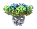
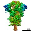
EMDB-27982:
Structure of the full-length IP3R1 channel determined at high Ca2+
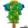
EMDB-27983:
Structure of the full-length IP3R1 channel determined in the presence of Calcium/IP3/ATP
PDB-8eaq:
Structure of the full-length IP3R1 channel determined at high Ca2+
PDB-8ear:
Structure of the full-length IP3R1 channel determined in the presence of Calcium/IP3/ATP

EMDB-24609:
Subnanometer in situ structure of the AcrAB-TolC efflux pump bound with MBX
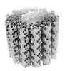
EMDB-26018:
The symmetry-released subpellicular microtubule map from detergent-extracted Toxoplasma cells
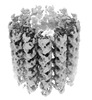
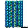
EMDB-26019:
Subpellicular microtubule from detergent-extract Toxoplasma gondii cells
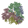
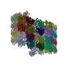
EMDB-26020:
The tubulin-based conoid from detergent-extract Toxoplasma gondii cells
PDB-7tnq:
The symmetry-released subpellicular microtubule map from detergent-extracted Toxoplasma cells
PDB-7tns:
Subpellicular microtubule from detergent-extract Toxoplasma gondii cells

PDB-7tnt:
The tubulin-based conoid from detergent-extract Toxoplasma gondii cells
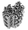
EMDB-26010:
The symmetrized subpellicular microtubule map from intact Toxoplasma gondii cells
EMDB-26006:
3D reconstruction of detergent-extract Toxoplasma gondii cells at the apical end
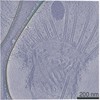
EMDB-26007:
Three-dimensional organization of the apical complex in Toxoplasma tachyzoites
EMDB-26008:
In situ average map of conoid with PCR from intact Toxoplasma gondii cells
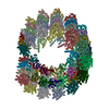
EMDB-26009:
The conoid segment from intact Toxoplasma gondii cells

EMDB-24287:
Cryo-ET platelet of WT mouse

EMDB-24288:
Cryo-ET platelet of AML mouse

EMDB-24289:
Cryo-ET platelet of pre-AML mouse

EMDB-24291:
Cryo-ET platelet of control mouse (1 week)

EMDB-24298:
Cryo-ET platelet of control mouse (3 week)

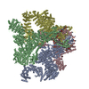
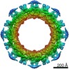
EMDB-24224:
Combined 3D structure of the isolated yeast NPC
EMDB-24225:
Low resolution, multi-part 3D structure of the isolated yeast NPC

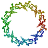
EMDB-24231:
Double nuclear outer ring from the isolated yeast NPC
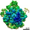
EMDB-24232:
Inner ring spoke from the isolated yeast NPC

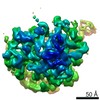
EMDB-24258:
Structure of the in situ yeast NPC
PDB-7n84:
Double nuclear outer ring from the isolated yeast NPC

PDB-7n85:
Inner ring spoke from the isolated yeast NPC

PDB-7n9f:
Structure of the in situ yeast NPC
EMDB-24129:
Classification of ribosome assembly intermediates
EMDB-24130:
Exploration of subtle structural variability of ribosome assembly intermediates
EMDB-24131:
Continuous movement of the central protuberance domain of 50S ribosome.
EMDB-24092:
Continuous movement of the pre-catalytic spliceosome, mode 1.
EMDB-24093:
Continuous movement of the pre-catalytic spliceosome, mode 2.
EMDB-24094:
Continuous movement of the pre-catalytic spliceosome, mode 1-2.
EMDB-24096:
Continuous movement of the pre-catalytic spliceosome, mode 1+2.
EMDB-24118:
Continuous motion of SARS-COV-2 spike protein receptor binding domain. Movement model 1.
EMDB-24119:
Continuous motion of SARS-COV-2 spike protein receptor binding domain. Movement model 2.
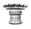
EMDB-23122:
Subtomogram average of the hexameric fungal plasma membrane proton pump Pma1 from protoplast membrane of Candida glabrata CBS138 strain
EMDB-23123:
Subtomogram average of the hexameric fungal plasma membrane proton pump Pma1 from protoplast membrane of Candida glabrata KH238 strain

EMDB-22079:
EM Structure of Full-Length Androgen Receptor and DNA Complex
EMDB-22080:
EM Structure of Full-Length Androgen Receptor Coactivator Complex
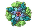
EMDB-20416:
Cryo-EM structure of the full-length Bacillus subtilis glyQS T-box riboswitch in complex with tRNA-Gly
PDB-6pom:
Cryo-EM structure of the full-length Bacillus subtilis glyQS T-box riboswitch in complex with tRNA-Gly

EMDB-0529:
Purified ribosome - A complete data processing workflow for CryoET and subtomogram averaging
ページ:
 wwPDBはEMDBデータモデルのバージョン3へ移行します
