 ムービー
ムービー コントローラー
 構造ビューア
 EMN検索について


-検索条件
-検索結果
検索 (著者・登録者: li & ht)の結果1,208件中、1から50件目までを表示しています
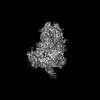
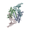
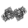
EMDB-44965: 
Sub-tomogram average of the RSV M lattice from native virions released from RSV-infected BEAS-2B cells cultured on EM grids

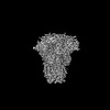
EMDB-44966:
Sub-tomogram average of a pair of RSV F trimers from native virions released from RSV-infected BEAS-2B cells cultured on EM grids

EMDB-44968:
Sub-tomogram average of two pairs of RSV F trimers from the surface of native virions released from RSV-infected BEAS-2B cells cultured on EM grids

EMDB-44969:
Sub-tomogram average of two pairs of RSV F trimers from the surface of native virions released from RSV-infected BEAS-2B cells cultured on EM grids

EMDB-44971:
Sub-tomogram average of two pairs of RSV F trimers from the surface of native virions released from RSV-infected BEAS-2B cells cultured on EM grids

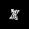
EMDB-45639:
Cryo-EM structure of in-vitro alpha-synuclein fibril
PDB-9ck3:
Cryo-EM structure of in-vitro alpha-synuclein fibril
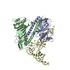
EMDB-44599:
Cryo-EM structure of the mammalian peptide transporter PepT2 bound to cefadroxil

EMDB-44600:
Cryo-EM structure of the mammalian peptide transporter PepT2 bound to amoxicillin
EMDB-44601:
Cryo-EM structure of the mammalian peptide transporter PepT2 bound to cloxacillin, pose 1
EMDB-44602:
Cryo-EM structure of the mammalian peptide transporter PepT2 bound to cloxacillin, pose 2

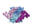
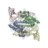
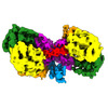
EMDB-39025:
Structure of HCoV-HKU1A spike in the functionally anchored-3up conformation with 3TMPRSS2

EMDB-39026:
Local structure of HCoV-HKU1A spike in complex with TMPRSS2 and glycan

EMDB-39036:
Structure of HCoV-HKU1C spike in the functionally anchored-1up conformation with 1TMPRSS2
EMDB-39037:
Structure of HCoV-HKU1C spike in the functionally anchored-2up conformation with 2TMPRSS2
EMDB-39038:
Structure of HCoV-HKU1C spike in the functionally anchored-3up conformation with 2TMPRSS2

EMDB-39039:
Structure of HCoV-HKU1C spike in the functionally anchored-3up conformation with 3TMPRSS2

EMDB-39040:
Local structure of HCoV-HKU1C spike in complex with TMPRSS2 and glycan
EMDB-39041:
Structure of HCoV-HKU1C spike in the inactive-closed conformation
EMDB-39042:
Structure of HCoV-HKU1C spike in the inactive-1up conformation

EMDB-39043:
Structure of HCoV-HKU1C spike in the inactive-2up conformation
EMDB-39044:
Structure of HCoV-HKU1C spike in the glycan-activated-closed conformation

EMDB-39045:
Structure of HCoV-HKU1C spike in the glycan-activated-1up conformation

EMDB-39046:
Structure of HCoV-HKU1C spike in the glycan-activated-2up conformation

EMDB-39047:
Structure of HCoV-HKU1C spike in the glycan-activated-3up conformation

EMDB-39048:
Local structure of HCoV-HKU1C spike in complex with glycan
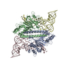
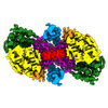
EMDB-16917:
Human apo pseudouridine synthase 3 (PUS3)

EMDB-16926:
Human pseudouridine synthase 3 and tRNA-Gln
EMDB-19830:
Human pseudouridine synthase 3 (PUS3 R116A mutant) and two tRNA-Gln

EMDB-19831:
Human pseudouridine synthase 3 (PUS3 R116A mutant) and one tRNA-Gln
EMDB-19832:
Human pseudouridine synthase 3 (PUS3 D118A mutant) and two tRNA-Arg
EMDB-19833:
Human pseudouridine synthase 3 (PUS3 D118A mutant) and two pre-tRNA-Arg
EMDB-19834:
Human pseudouridine synthase 3 (PUS3 R116A mutant)

EMDB-19835:
Human pseudouridine synthase 3 (PUS3 K119A mutant)
EMDB-19836:
Human pseudouridine synthase 3 (wild type)
PDB-8okd:
Human pseudouridine synthase 3 and tRNA-Gln
PDB-9enb:
Human pseudouridine synthase 3 (PUS3 R116A mutant) and two tRNA-Gln
PDB-9enc:
Human pseudouridine synthase 3 (PUS3 R116A mutant) and one tRNA-Gln
PDB-9ene:
Human pseudouridine synthase 3 (PUS3 D118A mutant) and two tRNA-Arg
PDB-9enf:
Human pseudouridine synthase 3 (PUS3 D118A mutant) and two pre-tRNA-Arg
PDB-9f9q:
Human apo pseudouridine synthase 3 (PUS3 D118A mutant)

EMDB-50580:
SOLIST cryo-tomogram of native left ventricle mouse heart muscle #1

EMDB-50582:
SOLIST native mouse heart muscle tomogram #2

EMDB-50358:
In vitro-induced genome-releasing intermediate of Rhodobacter microvirus Ebor computed with C5 symmetry
EMDB-43706:
Human DNA polymerase theta helicase domain in complex with ssDNA, dimer form
EMDB-43816:
Human DNA polymerase theta helicase domain in complex with AMP-PNP, dimer form
EMDB-43817:
Human DNA polymerase theta helicase domain dimer, apo-form

EMDB-43818:
Human DNA polymerase theta helicase domain tetramer, apo-form
EMDB-45217:
Human DNA polymerase theta helicase domain in microhomology annealed state 1, dimer form
EMDB-45218:
Human DNA polymerase theta helicase domain in microhomology annealed state 2, dimer form
ページ:
 wwPDBはEMDBデータモデルのバージョン3へ移行します
