 Movie
Movie Controller

[English] 日本語
 Yorodumi
- EMDB-50580: SOLIST cryo-tomogram of native left ventricle mouse heart muscle #1 -
+ Open data

- Basic information
| Entry |  | |||||||||
|---|---|---|---|---|---|---|---|---|---|---|
| Title | SOLIST cryo-tomogram of native left ventricle mouse heart muscle #1 | |||||||||
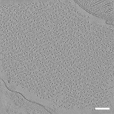
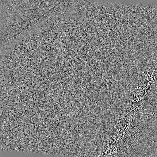
 Map data Map data | M. musculus heart muscle tomogram 1 (cross-section) | |||||||||
 Sample Sample |
| |||||||||
 Keywords Keywords | muscle / motor protein / thick filament / thin filament | |||||||||
| Biological species |  | |||||||||
| Method | electron tomography | |||||||||
 Authors Authors | Erdmann PS / Nguyen HTD / Perone G / Klena N / Vazzana R / Kaluthantrige Don F / Silva M / Sorrentino S / Swuec P / Leroux F ...Erdmann PS / Nguyen HTD / Perone G / Klena N / Vazzana R / Kaluthantrige Don F / Silva M / Sorrentino S / Swuec P / Leroux F / Kalebic N / Coscia F | |||||||||
| Funding support | 1 items
| |||||||||
 Citation Citation | Journal: Nat Methods / Year: 2024 Title: Serialized on-grid lift-in sectioning for tomography (SOLIST) enables a biopsy at the nanoscale. Authors: Ho Thuy Dung Nguyen / Gaia Perone / Nikolai Klena / Roberta Vazzana / Flaminia Kaluthantrige Don / Malan Silva / Simona Sorrentino / Paolo Swuec / Frederic Leroux / Nereo Kalebic / Francesca ...Authors: Ho Thuy Dung Nguyen / Gaia Perone / Nikolai Klena / Roberta Vazzana / Flaminia Kaluthantrige Don / Malan Silva / Simona Sorrentino / Paolo Swuec / Frederic Leroux / Nereo Kalebic / Francesca Coscia / Philipp S Erdmann /   Abstract: Cryo-focused ion beam milling has substantially advanced our understanding of molecular processes by opening windows into cells. However, applying this technique to complex samples, such as tissues, ...Cryo-focused ion beam milling has substantially advanced our understanding of molecular processes by opening windows into cells. However, applying this technique to complex samples, such as tissues, has presented considerable technical challenges. Here we introduce an innovative adaptation of the cryo-lift-out technique, serialized on-grid lift-in sectioning for tomography (SOLIST), addressing these limitations. SOLIST enhances throughput, minimizes ice contamination and improves sample stability for cryo-electron tomography. It thereby facilitates the high-resolution imaging of a wide range of specimens. We illustrate these advantages on reconstituted liquid-liquid phase-separated droplets, brain organoids and native tissues from the mouse brain, liver and heart. With SOLIST, cellular processes can now be investigated at molecular resolution directly in native tissue. Furthermore, our method has a throughput high enough to render cryo-lift-out a competitive tool for structural biology. This opens new avenues for unprecedented insights into cellular function and structure in health and disease, a 'biopsy at the nanoscale'. | |||||||||
| History |
|
- Structure visualization
| Supplemental images |
|---|
- Downloads & links
-EMDB archive
| Map data | emd_50580.map.gz | 235.9 MB |  EMDB map data format EMDB map data format | |
|---|---|---|---|---|
| Header (meta data) | emd-50580-v30.xmlemd-50580.xml | 11.6 KB 11.6 KB | Display Display | EMDB header |
| Images |  emd_50580.png emd_50580.png | 283.4 KB | ||
| Filedesc metadata | emd-50580.cif.gz | 4.5 KB | ||
| Archive directory |  http://ftp.pdbj.org/pub/emdb/structures/EMD-50580ftp://ftp.pdbj.org/pub/emdb/structures/EMD-50580 http://ftp.pdbj.org/pub/emdb/structures/EMD-50580ftp://ftp.pdbj.org/pub/emdb/structures/EMD-50580 | HTTPS FTP |
-Related structure data
| Related structure data | C: citing same article ( |
|---|


-Links
| EMDB pages | EMDB (EBI/PDBe) / EMDataResource |
|---|
-Map
| File | Download / File: emd_50580.map.gz / Format: CCP4 / Size: 256 MB / Type: IMAGE STORED AS FLOATING POINT NUMBER (4 BYTES) | ||||||||||||||||||||||||||||||||
|---|---|---|---|---|---|---|---|---|---|---|---|---|---|---|---|---|---|---|---|---|---|---|---|---|---|---|---|---|---|---|---|---|---|
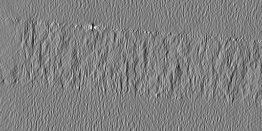
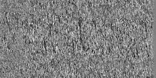
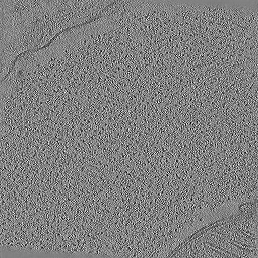
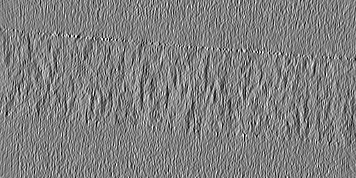
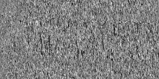
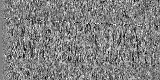
| Annotation | M. musculus heart muscle tomogram 1 (cross-section) | ||||||||||||||||||||||||||||||||
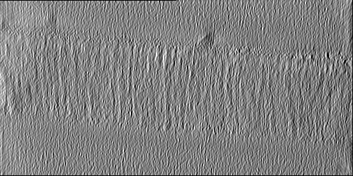
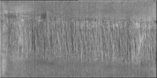
| Projections & slices | Image control
Images are generated by Spider. generated in cubic-lattice coordinate | ||||||||||||||||||||||||||||||||
| Voxel size | X=Y=Z: 15.624 Å | ||||||||||||||||||||||||||||||||
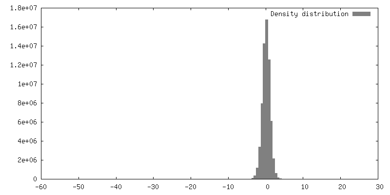
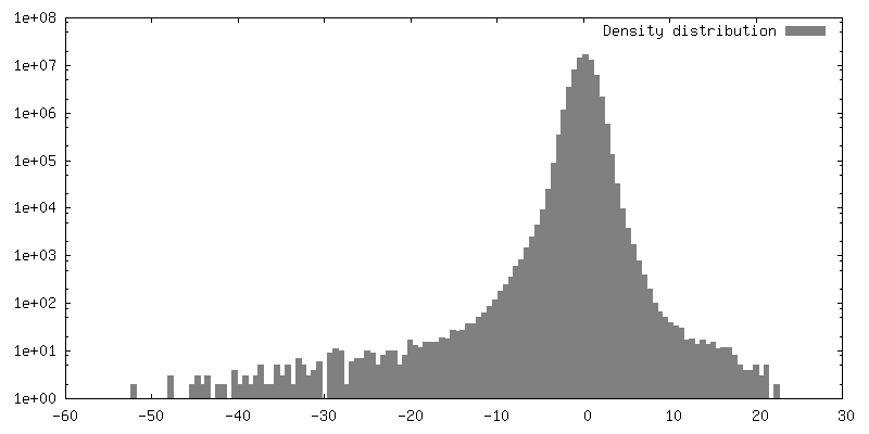
| Density |
| ||||||||||||||||||||||||||||||||
| Symmetry | Space group: 1 | ||||||||||||||||||||||||||||||||
| Details | EMDB XML:
|
 Z (Sec.)
 Y (Row.)
 X (Col.)


-Supplemental data
- Sample components
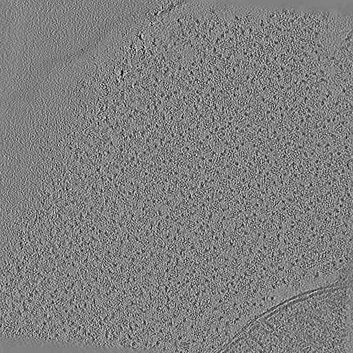
-Entire : In situ cryo-electron tomogram of native mouse heart muscle #1
| Entire | Name: In situ cryo-electron tomogram of native mouse heart muscle #1 |
|---|---|
| Components |
|
-Supramolecule #1: In situ cryo-electron tomogram of native mouse heart muscle #1
| Supramolecule | Name: In situ cryo-electron tomogram of native mouse heart muscle #1 type: cell / ID: 1 / Parent: 0 / Details: cross-section of thick and thin filaments |
|---|---|
| Source (natural) | Organism: |
-Experimental details
-Structure determination
 Processing Processing | electron tomography |
|---|---|
| Aggregation state | cell |
-Sample preparation
| Buffer | pH: 7.5 |
|---|---|
| Grid | Model: UltrAuFoil R2/2 / Material: GOLD / Mesh: 200 / Pretreatment - Type: GLOW DISCHARGE / Pretreatment - Time: 30 sec. Details: glow-discharged at 30 mA for 30 s (GloQube Plus Glow discharge system, Quorum Technologies) |
| Details | SOLIST lamella preparation of native mouse heart muscle |
| High pressure freezing | Instrument: OTHER Details: Before use, 3 mm type A (100 micrometer well) and type B planchettes were cleaned by sonication for 5 min, 30 s on, 30 s off, 40% Amplitude on a Branson SFX 550. Type B lids were polished ...Details: Before use, 3 mm type A (100 micrometer well) and type B planchettes were cleaned by sonication for 5 min, 30 s on, 30 s off, 40% Amplitude on a Branson SFX 550. Type B lids were polished with 1 micrometer lapping paper and coated with 0.1% soy lecithin dissolved in chloroform.. The value given for _em_high_pressure_freezing.instrument is Leica EM ICE. This is not in a list of allowed values {'LEICA EM PACT2', 'LEICA EM PACT', 'BAL-TEC HPM 010', 'EMS-002 RAPID IMMERSION FREEZER', 'LEICA EM HPM100', 'OTHER'} so OTHER is written into the XML file. |
| Cryo protectant | 20% dextran, 5% sucrose |
| Sectioning | Focused ion beam - Instrument: OTHER / Focused ion beam - Ion: OTHER / Focused ion beam - Voltage: 30 / Focused ion beam - Current: 0.03 / Focused ion beam - Duration: 60 / Focused ion beam - Temperature: 80 K / Focused ion beam - Initial thickness: 4000 / Focused ion beam - Final thickness: 200 Focused ion beam - Details: SOLIST procedure. The value given for _em_focused_ion_beam.instrument is Aquilos 2. This is not in a list of allowed values {'DB235', 'OTHER'} so OTHER is written into the XML file. |
- Electron microscopy
| Microscope | TFS KRIOS |
|---|---|
| Specialist optics | Energy filter - Name: TFS Selectris X / Energy filter - Slit width: 10 eV |
| Image recording | Film or detector model: FEI FALCON IV (4k x 4k) / Digitization - Dimensions - Width: 4096 pixel / Digitization - Dimensions - Height: 4096 pixel / Number real images: 41 / Average electron dose: 3.0 e/Å2 |
| Electron beam | Acceleration voltage: 300 kV / Electron source:  FIELD EMISSION GUN FIELD EMISSION GUN |
| Electron optics | Illumination mode: FLOOD BEAM / Imaging mode: BRIGHT FIELD / Nominal defocus max: 3.5 µm / Nominal defocus min: 1.5 µm |
| Sample stage | Specimen holder model: FEI TITAN KRIOS AUTOGRID HOLDER / Cooling holder cryogen: NITROGEN |
| Experimental equipment |  Model: Titan Krios / Image courtesy: FEI Company |
-Image processing
| Final reconstruction | Algorithm: BACK PROJECTION / Software - Name: eTomo (ver. 4.11.7) / Details: final tomogram denoised with CryoCare 2.1 / Number images used: 41 |
|---|
