 Movie
Movie Controller
 Structure viewers
 About EMN search


-Search query
-Search result
Showing all 41 items for (author: huang & yx)
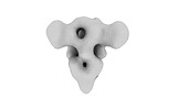
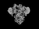
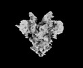
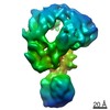
EMDB-39107: 
SARS-CoV-2 DMV nsp3-4 pore complex (full-pore)
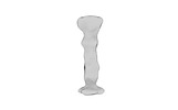
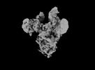
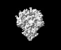
EMDB-39109:
SARS-CoV-2 DMV nsp3-4 pore complex (consensus-pore, C6 symmetry)
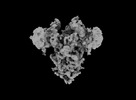
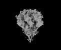
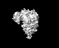
EMDB-39111:
SARS-CoV-2 DMV nsp3-4 pore complex (extended-pore)
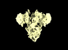
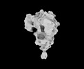
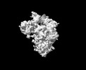
EMDB-39112:
SARS-CoV-2 DMV nsp3-4 pore complex (consensus-pore, C3 symmetry)
EMDB-39113:
SARS-CoV-2 DMV nsp3-4 pore complex (mini-pore)
EMDB-39159:
SARS-CoV-2 DMV nsp3-4 pore complex (full-length-pore)

EMDB-33990:
Cryo-EM structure of EBV gHgL-gp42 in complex with mAbs 3E8 and 5E3 (localized refinement)

EMDB-33992:
Cryo-EM structure of EBV gHgL-gp42 in complex with mAb 10E4 (localized refinement)

EMDB-33993:
Cryo-EM density map of EBV gHgL-gp42 in complex with four mAbs 5E3, 3E8, 6H2 and 10E4

EMDB-33994:
Cryo-EM structure of EBV gHgL-gp42 in complex with mAb 6H2 (localized refinement)
EMDB-33102:
Cryo-EM structure of EBV glycoprotein complex gHgL-gp42 bound by a neutralizing antibody 6H2
EMDB-32329:
Cryo-EM map of PEDV (Pintung 52) S protein with all three protomers in the D0-down conformation determined in situ on intact viral particles.
EMDB-32332:
Subtomogram averaging of PEDV (Pintung 52) S protein with all three protomers in the D0-down conformation determined in situ on intact viral particles.
EMDB-32333:
Subtomogram averaging of PEDV (Pintung 52) S protein with one protomer in the D0-up conformation and two protomers in the D0-down conformation, determined in situ on intact viral particles
EMDB-32337:
Subtomogram averaging of PEDV (Pintung 52) S protein with two protomers in the D0-up conformation and one protomer in the D0-down conformation, determined in situ on intact viral particles.
EMDB-32338:
Cryo-EM map of PEDV S protein with one protomer in the D0-up conformation while the other two in the D0-down conformation
EMDB-32339:
Subtomogram averaging of PEDV (Pintung 52) S protein with all three protomers in the D0-up conformation determined in situ on intact viral particles.
EMDB-32340:
Subtomogram averaging of PEDV (Pintung 52) S protein in the postfusion form determined in situ on intact viral particles.
EMDB-33646:
Cryo-EM map of IPEC-J2 cell-derived PEDV PT52 S protein with three D0-up
EMDB-33647:
Cryo-EM map of IPEC-J2 cell-derived PEDV PT52 S protein one D0-down and two D0-up
EMDB-33648:
Symmetry-expanded and locally refined protomer structure of IPEC-J2 cell-derived PEDV PT52 S with a CTD-close conformation
EMDB-33649:
Symmetry-expanded and locally refined protomer structure of IPEC-J2 cell-derived PEDV PT52 S with a CTD-open conformation
EMDB-33700:
Cryo-EM map of HEK293F cell-derived PEDV PT52 S protein with three D0-down
EMDB-33701:
Cryo-EM map of HEK293F cell-derived PEDV PT52 S protein one D0-up and two D0-down
EMDB-33702:
Cryo-EM map of HEK293F cell-derived PEDV PT52 S protein with three D0-up
EMDB-33703:
Cryo-EM map of HEK293F cell-derived PEDV PT52 S T326I with three D0-down
EMDB-33704:
Cryo-EM map of HEK293F cell-derived PEDV PT52 S T326I one D0-up and two D0-down
EMDB-33705:
Cryo-EM map of HEK293F cell-derived PEDV PT52 S T326I one D0-down and two D0-up
EMDB-33706:
Cryo-EM map of HEK293F cell-derived PEDV PT52 S T326I with three D0-up

EMDB-32557:
SARS-CoV-2 Omicron S-open
EMDB-32170:
SARS-CoV-2 Beta variant spike protein in transition state
EMDB-31146:
Coupling of N7-methyltransferase and 3'-5' exoribonuclease with SARS-CoV-2 polymerase reveals mechanisms for capping and proofreading
EMDB-31138:
Co-transcriptional capping machineries in SARS-CoV-2 RTC: Coupling of N7-methyltransferase and 3'-5' exoribonuclease with polymerase reveals mechanisms for capping and proofreading

EMDB-21249:
Cryo-EM structure of an activated VIP1 receptor-G protein complex
EMDB-20417:
Cryo-EM reconstruction of the mature chimeric BinJV/ZIKV-prME virion
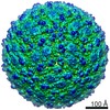
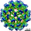
EMDB-20438:
Cryo-EM reconstruction of the mature chimeric BinJV/ZIKV-prME virion in complex with Fab C8
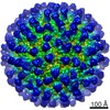
EMDB-20439:
Cryo-EM reconstruction of the immature chimeric BinJV/ZIKV-prME virion
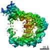
EMDB-20416:
Cryo-EM structure of the full-length Bacillus subtilis glyQS T-box riboswitch in complex with tRNA-Gly
EMDB-9694:
The complete structure of yeast COMPASS
EMDB-2870:
Cryo-EM structure of the multimeric complex between Dark and Dronc-CARD.
EMDB-2871:
Cryo-EM structure of the Dark apoptosome.
 wwPDB to switch to version 3 of the EMDB data model
