 Movie
Movie Controller


+ Open data

- Basic information
| Entry | Database: EMDB / ID: EMD-2871 | |||||||||
|---|---|---|---|---|---|---|---|---|---|---|
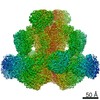
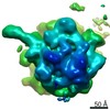
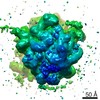
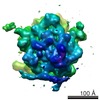
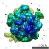
| Title | Cryo-EM structure of the Dark apoptosome. | |||||||||
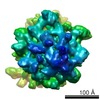
 Map data Map data | Reconstruction of Dark apoptosome | |||||||||
 Sample Sample |
| |||||||||
 Keywords Keywords | Cryo-EM / apoptosome / single particle analysis | |||||||||
| Function / homology |  Function and homology information Function and homology informationnegative regulation of humoral immune response / positive regulation of glial cell apoptotic process / Formation of apoptosome / salivary gland histolysis / melanization defense response / : / Regulation of the apoptosome activity / Activation of caspases through apoptosome-mediated cleavage / compound eye retinal cell programmed cell death / central nervous system formation ...negative regulation of humoral immune response / positive regulation of glial cell apoptotic process / Formation of apoptosome / salivary gland histolysis / melanization defense response / : / Regulation of the apoptosome activity / Activation of caspases through apoptosome-mediated cleavage / compound eye retinal cell programmed cell death / central nervous system formation / positive regulation of apoptotic process involved in development / sperm individualization / apoptosome / autophagic cell death / chaeta development / Neutrophil degranulation / L-methionine cycle / CARD domain binding / programmed cell death / triglyceride homeostasis / dendrite morphogenesis / response to starvation / response to gamma radiation / neuron cellular homeostasis / ADP binding / apoptotic process / structural molecule activity / ATP binding / identical protein binding Similarity search - Function | |||||||||
| Biological species |  | |||||||||
| Method | single particle reconstruction / cryo EM / negative staining / Resolution: 4.0 Å | |||||||||
 Authors Authors | Pang YX / Bai XC / Hao Q / Yan CY / Chen ZQ / Wang JW / Scheres SHW / Shi YG | |||||||||
 Citation Citation | Journal: Genes Dev / Year: 2015 Title: Structure of the apoptosome: mechanistic insights into activation of an initiator caspase from Drosophila. Authors: Yuxuan Pang / Xiao-chen Bai / Chuangye Yan / Qi Hao / Zheqin Chen / Jia-Wei Wang / Sjors H W Scheres / Yigong Shi /   Abstract: Apoptosis is executed by a cascade of caspase activation. The autocatalytic activation of an initiator caspase, exemplified by caspase-9 in mammals or its ortholog, Dronc, in fruit flies, is ...Apoptosis is executed by a cascade of caspase activation. The autocatalytic activation of an initiator caspase, exemplified by caspase-9 in mammals or its ortholog, Dronc, in fruit flies, is facilitated by a multimeric adaptor complex known as the apoptosome. The underlying mechanism by which caspase-9 or Dronc is activated by the apoptosome remains unknown. Here we report the electron cryomicroscopic (cryo-EM) structure of the intact apoptosome from Drosophila melanogaster at 4.0 Å resolution. Analysis of the Drosophila apoptosome, which comprises 16 molecules of the Dark protein (Apaf-1 ortholog), reveals molecular determinants that support the assembly of the 2.5-MDa complex. In the absence of dATP or ATP, Dronc zymogen potently induces formation of the Dark apoptosome, within which Dronc is efficiently activated. At 4.1 Å resolution, the cryo-EM structure of the Dark apoptosome bound to the caspase recruitment domain (CARD) of Dronc (Dronc-CARD) reveals two stacked rings of Dronc-CARD that are sandwiched between two octameric rings of the Dark protein. The specific interactions between Dronc-CARD and both the CARD and the WD40 repeats of a nearby Dark protomer are indispensable for Dronc activation. These findings reveal important mechanistic insights into the activation of initiator caspase by the apoptosome. | |||||||||
| History |
|
- Structure visualization
| Movie |
Movie viewer |
|---|---|
| Structure viewer | EM map: SurfViewMolmilJmol/JSmol |
| Supplemental images |

- Downloads & links
-EMDB archive
| Map data | emd_2871.map.gz | 115.7 MB | EMDB map data format | |
|---|---|---|---|---|
| Header (meta data) | emd-2871-v30.xmlemd-2871.xml | 9.9 KB 9.9 KB | Display Display | EMDB header |
| Images | 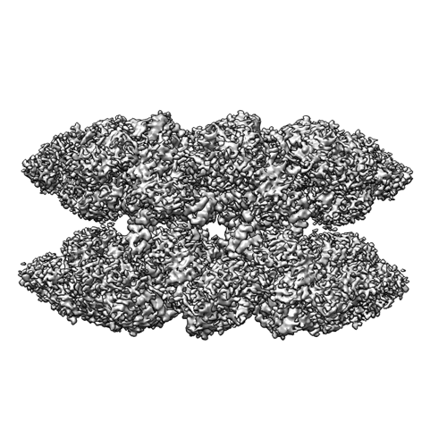 EMD_2871.png EMD_2871.png | 200.6 KB | ||
| Archive directory |  http://ftp.pdbj.org/pub/emdb/structures/EMD-2871ftp://ftp.pdbj.org/pub/emdb/structures/EMD-2871 http://ftp.pdbj.org/pub/emdb/structures/EMD-2871ftp://ftp.pdbj.org/pub/emdb/structures/EMD-2871 | HTTPS FTP |
-Related structure data
| Related structure data |  3j9lMC 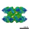 2870C 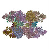 3j9kC M: atomic model generated by this map C: citing same article ( |
|---|---|
| Similar structure data |
-Links
| EMDB pages | EMDB (EBI/PDBe) / EMDataResource |
|---|---|
| Related items in Molecule of the Month |


-Map
| File | Download / File: emd_2871.map.gz / Format: CCP4 / Size: 122.1 MB / Type: IMAGE STORED AS FLOATING POINT NUMBER (4 BYTES) | ||||||||||||||||||||||||||||||||||||||||||||||||||||||||||||||||||||
|---|---|---|---|---|---|---|---|---|---|---|---|---|---|---|---|---|---|---|---|---|---|---|---|---|---|---|---|---|---|---|---|---|---|---|---|---|---|---|---|---|---|---|---|---|---|---|---|---|---|---|---|---|---|---|---|---|---|---|---|---|---|---|---|---|---|---|---|---|---|
| Annotation | Reconstruction of Dark apoptosome | ||||||||||||||||||||||||||||||||||||||||||||||||||||||||||||||||||||
| Projections & slices | Image control
Images are generated by Spider. | ||||||||||||||||||||||||||||||||||||||||||||||||||||||||||||||||||||
| Voxel size | X=Y=Z: 1.34 Å | ||||||||||||||||||||||||||||||||||||||||||||||||||||||||||||||||||||
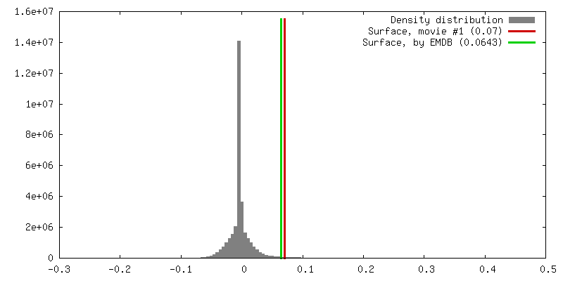
| Density |
| ||||||||||||||||||||||||||||||||||||||||||||||||||||||||||||||||||||
| Symmetry | Space group: 1 | ||||||||||||||||||||||||||||||||||||||||||||||||||||||||||||||||||||
| Details | EMDB XML:
CCP4 map header:
| ||||||||||||||||||||||||||||||||||||||||||||||||||||||||||||||||||||
 Z (Sec.)
 Y (Row.)
 X (Col.)


-Supplemental data
- Sample components
-Entire : Dark apoptosome
| Entire | Name: Dark apoptosome |
|---|---|
| Components |
|
-Supramolecule #1000: Dark apoptosome
| Supramolecule | Name: Dark apoptosome / type: sample / ID: 1000 / Oligomeric state: homo 16 mers / Number unique components: 1 |
|---|---|
| Molecular weight | Experimental: 2.5 MDa / Theoretical: 2.5 MDa |
-Macromolecule #1: Dark apoptosome
| Macromolecule | Name: Dark apoptosome / type: protein_or_peptide / ID: 1 / Recombinant expression: Yes |
|---|---|
| Source (natural) | Organism: |
| Molecular weight | Experimental: 2.5 MDa / Theoretical: 2.5 MDa |
| Recombinant expression | Organism: |
-Experimental details
-Structure determination
| Method | negative staining, cryo EM |
|---|---|
 Processing Processing | single particle reconstruction |
| Aggregation state | particle |
-Sample preparation
| Concentration | 0.15 mg/mL |
|---|---|
| Buffer | pH: 8 / Details: 25mM Tris, 150mM NaCl, 5mM dithiothreitol |
| Staining | Type: NEGATIVE Details: Grids were blotted for 2 seconds and flash frozen in liquid ethane using an FEI Vitrobot. |
| Grid | Details: Samples were placed on glow-discharged holey carbon grids (Quantifoil CuR2/2), on which a home-made continuous carbon film had previously been deposited. |
| Vitrification | Cryogen name: ETHANE / Chamber humidity: 100 % / Chamber temperature: 85 K / Instrument: FEI VITROBOT MARK II / Method: Blot for 2 seconds before plunging |
- Electron microscopy
| Microscope | FEI POLARA 300 |
|---|---|
| Temperature | Min: 80 K / Max: 90 K / Average: 85 K |
| Date | Nov 17, 2013 |
| Image recording | Category: CCD / Film or detector model: FEI FALCON II (4k x 4k) / Digitization - Sampling interval: 14 µm / Number real images: 693 / Average electron dose: 28 e/Å2 Details: 16 video frames were recorded in 1s by FalconII detector. |
| Electron beam | Acceleration voltage: 300 kV / Electron source:  FIELD EMISSION GUN FIELD EMISSION GUN |
| Electron optics | Calibrated magnification: 104748 / Illumination mode: FLOOD BEAM / Imaging mode: BRIGHT FIELD / Cs: 2 mm / Nominal defocus max: 6.6 µm / Nominal defocus min: 1.6 µm / Nominal magnification: 78000 |
| Sample stage | Specimen holder model: GATAN LIQUID NITROGEN |
| Experimental equipment |  Model: Tecnai Polara / Image courtesy: FEI Company |
-Image processing
| CTF correction | Details: Each particle |
|---|---|
| Final reconstruction | Applied symmetry - Point group: D8 (2x8 fold dihedral) / Resolution.type: BY AUTHOR / Resolution: 4.0 Å / Resolution method: OTHER / Software - Name: CTFFIND3, RELION Details: To correct for beam-induced movements, the 16 video frames for each micrograph were first aligned using whole-image motion correction. Second, particle based beam-induced movement correction ...Details: To correct for beam-induced movements, the 16 video frames for each micrograph were first aligned using whole-image motion correction. Second, particle based beam-induced movement correction was performed using statistical movie processing in RELION. Number images used: 9354 |
